

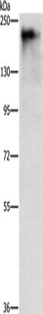
Gel: 8%SDS-PAGE, Lysate: 40 ug, Lane: A431 cells, Primary antibody: CSB-PA052246(ATAD5 Antibody) at dilution 1/400, Secondary antibody: Goat anti rabbit IgG at 1/8000 dilution, Exposure time: 15 seconds
Gel: 8%SDS-PAGE, Lysate: 40 ug, Lane: A431 cells, Primary antibody: CSB-PA052246(ATAD5 Antibody) at dilution 1/400, Secondary antibody: Goat anti rabbit IgG at 1/8000 dilution, Exposure time: 15 seconds
ATAD5 Antibody
CSB-PA052246
ApplicationsWestern Blot, ELISA
Product group Antibodies
ReactivityHuman
TargetATAD5
Overview
- SupplierCusabio
- Product NameATAD5 Antibody
- Delivery Days Customer20
- ApplicationsWestern Blot, ELISA
- CertificationResearch Use Only
- ClonalityPolyclonal
- ConjugateUnconjugated
- Gene ID79915
- Target nameATAD5
- Target descriptionATPase family AAA domain containing 5
- Target synonymsC17orf41, ELG1, FRAG1, ATPase family AAA domain-containing protein 5, chromosome fragility associated gene 1, chromosome fragility-associated gene 1 protein, enhanced level of genomic instability 1 homolog
- HostRabbit
- IsotypeIgG
- Protein IDQ96QE3
- Protein NameATPase family AAA domain-containing protein 5
- Scientific DescriptionATAD5 (ATPase family, AAA domain containing 5), also known as chromosome fragility-associated gene 1 protein, FRAG1 or ELG1, is a 1,844 amino acid nuclear protein that is involved in the DNA damage response and belongs to the AAA ATPase family. Existing as two alternatively spliced isoforms, ATAD5 interacts with Rad9 in growing cells where it assists in interactions between Rad9 and Bcl-2.
- ReactivityHuman
- Storage Instruction-20°C or -80°C
- UNSPSC41116161
Related products

Product group Antibodies

Goat anti-ATAD5 / FRAG1EB07634
ApplicationsFlow Cytometry, ImmunoFluorescence, ELISA
ReactivityHuman
TargetATAD5
- SizePrice

Product group Antibodies
Anti-ATAD5 AntibodyHPA066823
ApplicationsImmunoCytoChemistry
ReactivityHuman
TargetATAD5
- SizePrice
Product group Antibodies

ATAD5 / ELG1 AntibodyLS-C406232
ApplicationsWestern Blot, ELISA
ReactivityHuman
TargetATAD5
- SizePrice
![Western blot using GeneTex Affinity Purified anti-Elg1 antibody (GTX25429) shows detection of a band ~120 kDa corresponding to human Elg1 in various cell lysates. Lanes contain ~ 5ug of HeLa nuclear extract (1), HeLa (2), A431 (3), Jurkat (4) and HEK293 (5) whole cell lysates. After SDS-PAGE, transfer and blocking, the membrane was probed with the primary antibody diluted to 1:500. The membrane was then washed and reacted with a HRP conjugated Gt-a-Rabbit IgG [H&L] MX followed by ECL detection using a 2 m exposure time. The expected molecular weight of Elg1 is 120kDa according to Kanellis P et al. 2003, although the predicted molecular weight is 207 kDa. The 50kD bands in Jurkat and 293 cell lysates are probably cross-reaction with other proteins. Both the 120 kDa and 50 kDa bands are not observed when antibody is pre-incubated with peptide (data not shown](https://www.genetex.com/upload/website/prouct_img/normal/GTX25429/GTX25429_20160330_WB_w_23060722_890.webp)
Product group Antibodies
ATAD5 antibodyGTX25429
ApplicationsWestern Blot, ELISA
ReactivityHuman
TargetATAD5
- SizePrice
Product group Antibodies
ATAD5 Polyclonal AntibodyBS-7656R
ApplicationsImmunoFluorescence, Western Blot, ELISA, ImmunoCytoChemistry, ImmunoHistoChemistry, ImmunoHistoChemistry Frozen, ImmunoHistoChemistry Paraffin
ReactivityBovine, Canine, Chicken, Human, Mouse, Porcine, Rabbit, Rat
TargetATAD5
- SizePrice