

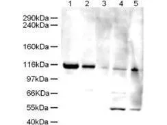
Western blot using GeneTex Affinity Purified anti-Elg1 antibody (GTX25429) shows detection of a band ~120 kDa corresponding to human Elg1 in various cell lysates. Lanes contain ~ 5ug of HeLa nuclear extract (1), HeLa (2), A431 (3), Jurkat (4) and HEK293 (5) whole cell lysates. After SDS-PAGE, transfer and blocking, the membrane was probed with the primary antibody diluted to 1:500. The membrane was then washed and reacted with a HRP conjugated Gt-a-Rabbit IgG [H&L] MX followed by ECL detection using a 2 m exposure time. The expected molecular weight of Elg1 is 120kDa according to Kanellis P et al. 2003, although the predicted molecular weight is 207 kDa. The 50kD bands in Jurkat and 293 cell lysates are probably cross-reaction with other proteins. Both the 120 kDa and 50 kDa bands are not observed when antibody is pre-incubated with peptide (data not shown

Western blot using GeneTex Affinity Purified anti-Elg1 antibody (GTX25429) shows detection of a band ~120 kDa corresponding to human Elg1 in various cell lysates. Lanes contain ~ 5ug of HeLa nuclear extract (1), HeLa (2), A431 (3), Jurkat (4) and HEK293 (5) whole cell lysates. After SDS-PAGE, transfer and blocking, the membrane was probed with the primary antibody diluted to 1:500. The membrane was then washed and reacted with a HRP conjugated Gt-a-Rabbit IgG [H&L] MX followed by ECL detection using a 2 m exposure time. The expected molecular weight of Elg1 is 120kDa according to Kanellis P et al. 2003, although the predicted molecular weight is 207 kDa. The 50kD bands in Jurkat and 293 cell lysates are probably cross-reaction with other proteins. Both the 120 kDa and 50 kDa bands are not observed when antibody is pre-incubated with peptide (data not shown
ATAD5 antibody
GTX25429
ApplicationsWestern Blot, ELISA
Product group Antibodies
ReactivityHuman
TargetATAD5
Overview
- SupplierGeneTex
- Product NameATAD5 antibody
- Delivery Days Customer9
- Application Supplier NoteWB: 1:500-1:2000. ELISA: 1:3000-1:15000. *Optimal dilutions/concentrations should be determined by the researcher.Not tested in other applications.
- ApplicationsWestern Blot, ELISA
- CertificationResearch Use Only
- ClonalityPolyclonal
- Concentration0.5 mg/ml
- ConjugateUnconjugated
- Gene ID79915
- Target nameATAD5
- Target descriptionATPase family AAA domain containing 5
- Target synonymsC17orf41, ELG1, FRAG1, ATPase family AAA domain-containing protein 5, chromosome fragility associated gene 1, chromosome fragility-associated gene 1 protein, enhanced level of genomic instability 1 homolog
- HostRabbit
- IsotypeIgG
- Protein IDQ96QE3
- Protein NameATPase family AAA domain-containing protein 5
- Scientific DescriptionElg1 may be part of an alternative RFC complex involved ensuring replication fidelity.
- ReactivityHuman
- Storage Instruction-20°C or -80°C,2°C to 8°C
- UNSPSC41116161
Datasheet
Related products

Product group Antibodies

Goat anti-ATAD5 / FRAG1EB07634
ApplicationsFlow Cytometry, ImmunoFluorescence, ELISA
ReactivityHuman
TargetATAD5
- SizePrice
Product group Antibodies
ATAD5 AntibodyCSB-PA052246
ApplicationsWestern Blot, ELISA
ReactivityHuman
TargetATAD5
- SizePrice

Product group Antibodies
Anti-ATAD5 AntibodyHPA066823
ApplicationsImmunoCytoChemistry
ReactivityHuman
TargetATAD5
- SizePrice
Product group Antibodies

ATAD5 / ELG1 AntibodyLS-C406232
ApplicationsWestern Blot, ELISA
ReactivityHuman
TargetATAD5
- SizePrice
Product group Antibodies
ATAD5 Polyclonal AntibodyBS-7656R
ApplicationsImmunoFluorescence, Western Blot, ELISA, ImmunoCytoChemistry, ImmunoHistoChemistry, ImmunoHistoChemistry Frozen, ImmunoHistoChemistry Paraffin
ReactivityBovine, Canine, Chicken, Human, Mouse, Porcine, Rabbit, Rat
TargetATAD5
- SizePrice