

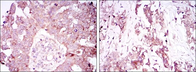
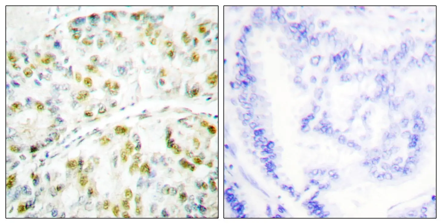
IHC-P analysis of ovarian cancer tissue (left) and lung cancer tissue (right) using GTX80399 Ataxin 1 antibody [2F5].
![ICC/IF analysis of NTERA-2 cells using GTX80399 Ataxin 1 antibody [2F5]. Green : Ataxin 1 Blue: DRAQ5 fluorescent DNA dye Red: Actin filaments](https://www.genetex.com/upload/website/prouct_img/normal/GTX80399/GTX80399_20170912_ICCIF_w_23061322_740.webp "ICC/IF analysis of NTERA-2 cells using GTX80399 Ataxin 1 antibody [2F5]. Green : Ataxin 1 Blue: DRAQ5 fluorescent DNA dye Red: Actin filaments")
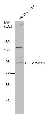
![WB analysis of HEK293 (1) and ATXN1(AA: 645-815)-hIgGFc transfected HEK293 (2) cell lysate using GTX80399 Ataxin 1 antibody [2F5].](https://www.genetex.com/upload/website/prouct_img/normal/GTX80399/GTX80399_20170912_WB_w_23061322_895.webp "WB analysis of HEK293 (1) and ATXN1(AA: 645-815)-hIgGFc transfected HEK293 (2) cell lysate using GTX80399 Ataxin 1 antibody [2F5].")
![FACS analysis of Jurkat cells using GTX80399 Ataxin 1 antibody [2F5]. Green : Ataxin 1 Purple : negative control](https://www.genetex.com/upload/website/prouct_img/normal/GTX80399/GTX80399_20170912_FACS_w_23061322_219.webp "FACS analysis of Jurkat cells using GTX80399 Ataxin 1 antibody [2F5]. Green : Ataxin 1 Purple : negative control")
IHC-P analysis of ovarian cancer tissue (left) and lung cancer tissue (right) using GTX80399 Ataxin 1 antibody [2F5].
Ataxin 1 antibody [2F5]
GTX80399
ApplicationsFlow Cytometry, ImmunoFluorescence, Western Blot, ELISA, ImmunoCytoChemistry, ImmunoHistoChemistry, ImmunoHistoChemistry Paraffin
Product group Antibodies
ReactivityHuman
TargetATXN1
Overview
- SupplierGeneTex
- Product NameAtaxin 1 antibody [2F5]
- Delivery Days Customer9
- Application Supplier NoteWB: 1/500 - 1/2000. ICC/IF: 1/200 - 1/1000. IHC-P: 1/200 - 1/1000. FCM: 1/200 - 1/400. ELISA: 1/10000. *Optimal dilutions/concentrations should be determined by the researcher.Not tested in other applications.
- ApplicationsFlow Cytometry, ImmunoFluorescence, Western Blot, ELISA, ImmunoCytoChemistry, ImmunoHistoChemistry, ImmunoHistoChemistry Paraffin
- CertificationResearch Use Only
- ClonalityMonoclonal
- Clone ID2F5
- ConjugateUnconjugated
- Gene ID6310
- Target nameATXN1
- Target descriptionataxin 1
- Target synonymsATX1, D6S504E, SCA1, ataxin-1, alternative ataxin1, spinocerebellar ataxia type 1 protein
- HostMouse
- IsotypeIgG1
- Protein IDP54253
- Protein NameAtaxin-1
- Scientific DescriptionThe autosomal dominant cerebellar ataxias (ADCA) are a heterogeneous group of neurodegenerative disorders characterized by progressive degeneration of the cerebellum, brain stem and spinal cord. Clinically, ADCA has been divided into three groups: ADCA types I-III. ADCAI is genetically heterogeneous, with five genetic loci, designated spinocerebellar ataxia (SCA) 1, 2, 3, 4 and 6, being assigned to five different chromosomes. ADCAII, which always presents with retinal degeneration (SCA7), and ADCAIII often referred to as the pure cerebellar syndrome (SCA5), are most likely homogeneous disorders. Several SCA genes have been cloned and shown to contain CAG repeats in their coding regions. ADCA is caused by the expansion of the CAG repeats, producing an elongated polyglutamine tract in the corresponding protein. The expanded repeats are variable in size and unstable, usually increasing in size when transmitted to successive generations. The function of the ataxins is not known. This loc
- ReactivityHuman
- Storage Instruction-20°C or -80°C,2°C to 8°C
- UNSPSC41116161
Datasheet
Related products
Product group Antibodies
Anti-Ataxin 1 AntibodyA96343
ApplicationsImmunoFluorescence, ELISA, ImmunoHistoChemistry
ReactivityHuman, Mouse
- SizePrice
Product group Antibodies
Anti-ATXN1 Antibody101-10137
ApplicationsWestern Blot, ELISA
TargetATXN1
- SizePrice
Product group Antibodies
ApplicationsImmunoFluorescence, Western Blot, ELISA, ImmunoCytoChemistry, ImmunoHistoChemistry, ImmunoHistoChemistry Frozen, ImmunoHistoChemistry Paraffin
ReactivityBovine, Canine, Chicken, Equine, Human, Mouse, Porcine, Rabbit, Rat, Sheep
TargetATXN1
- SizePrice
Product group Antibodies
ATXN1 AntibodyCSB-PA000943
ApplicationsImmunoFluorescence, Western Blot, ELISA, ImmunoHistoChemistry
ReactivityHuman, Mouse
TargetATXN1
- SizePrice
Product group Antibodies
ApplicationsImmunoPrecipitation, Western Blot, ImmunoCytoChemistry, ImmunoHistoChemistry
ReactivityMouse, Rat
TargetATXN1
- SizePrice
Product group Antibodies

ATXN1 / SCA1 AntibodyLS-C401437
ApplicationsELISA, ImmunoHistoChemistry
ReactivityHuman, Mouse, Rat
TargetATXN1
- SizePrice
Product group Antibodies
Ataxin 1 antibodyGTX87190
ApplicationsImmunoFluorescence, ImmunoCytoChemistry, ImmunoHistoChemistry, ImmunoHistoChemistry Paraffin
ReactivityHuman, Mouse
TargetATXN1
- SizePrice

Product group Antibodies
Anti-ATXN1 AntibodyHPA008335
ApplicationsImmunoHistoChemistry
ReactivityHuman
TargetATXN1
- SizePrice
Product group Antibodies
Ataxin 1 antibodyGTX130595
ApplicationsWestern Blot
ReactivityHuman, Mouse
TargetATXN1
- SizePrice