

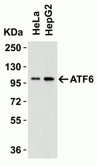
WB analysis of various whole cell lysates using GTX15457 AFT6 antibody. Dilution : 1μg/ml Loading : 15μg per lane
 and ATF6 knockout (KO) HeLa cell lysates using GTX15457 AFT6 antibody. Dilution : 0.5μg/ml Loading : 15μg per lane")


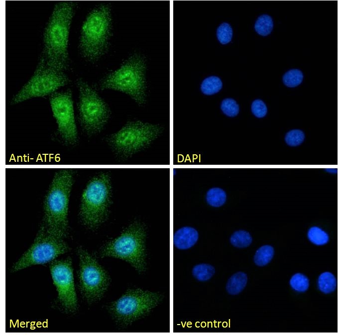
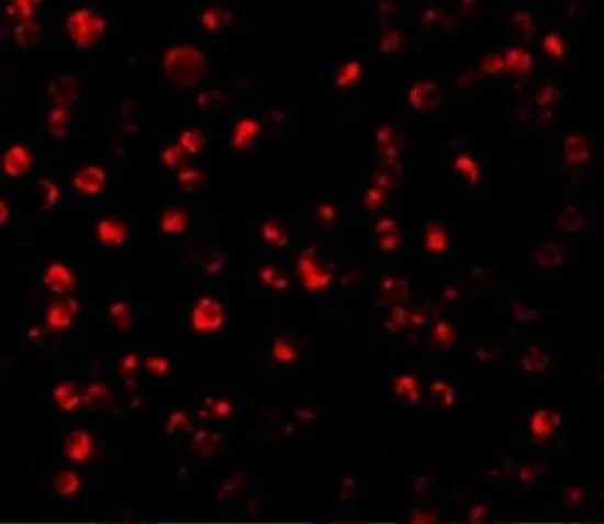
. Dilution : 5μg/ml")
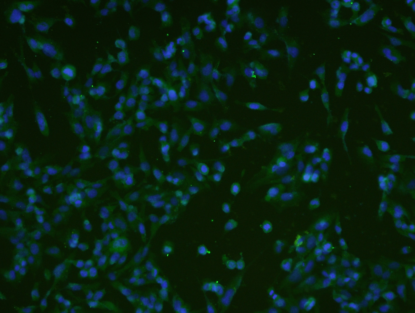
. Dilution : 5μg/ml")
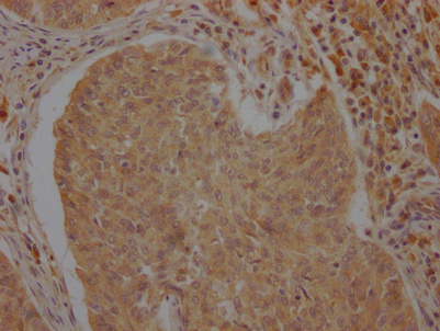
. Dilution : 5μg/ml")
WB analysis of various whole cell lysates using GTX15457 AFT6 antibody. Dilution : 1μg/ml Loading : 15μg per lane
ATF6 antibody
GTX15457
ApplicationsWestern Blot, ELISA, ImmunoHistoChemistry, ImmunoHistoChemistry Paraffin
Product group Antibodies
ReactivityHuman, Mouse, Rat
TargetATF6
Overview
- SupplierGeneTex
- Product NameATF6 antibody
- Delivery Days Customer9
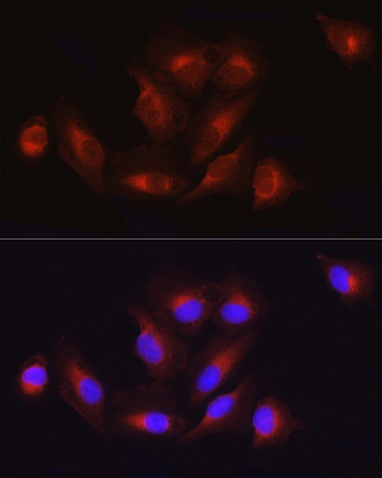
- Application Supplier NoteWB: 0.5-2 microg/mL. IHC-P: 5 microg/mL. *Optimal dilutions/concentrations should be determined by the researcher.Not tested in other applications.
- ApplicationsWestern Blot, ELISA, ImmunoHistoChemistry, ImmunoHistoChemistry Paraffin
- CertificationResearch Use Only
- ClonalityPolyclonal
- Concentration1 mg/ml
- ConjugateUnconjugated
- Gene ID22926
- Target nameATF6
- Target descriptionactivating transcription factor 6
- Target synonymsACHM7, ATF6A, ATP6alpha, cyclic AMP-dependent transcription factor ATF-6 alpha, cAMP-dependent transcription factor ATF-6 alpha
- HostRabbit
- IsotypeIgG
- Protein IDP18850
- Protein NameCyclic AMP-dependent transcription factor ATF-6 alpha
- Scientific DescriptionATF6 is an endoplasmic reticulum (ER) stress-regulated transmembrane transcription factor that activates the transcription of ER molecules.[supplied by OMIM]
- ReactivityHuman, Mouse, Rat
- Storage Instruction-20°C or -80°C,2°C to 8°C
- UNSPSC41116161
References
- ATF6alpha contributes to rheumatoid arthritis by inducing inflammatory cytokine production and apoptosis resistance.Read this paper
Datasheet
Related products
Product group Antibodies
Anti-ATF6 AntibodyA285927
ApplicationsFlow Cytometry, ImmunoFluorescence, ELISA
ReactivityHuman
- SizePrice

Product group Antibodies
Anti-ATF6 Antibody Picoband(r)A00655-3-CARRIER-FREE
ApplicationsFlow Cytometry, ImmunoFluorescence, Western Blot, ELISA, ImmunoCytoChemistry
ReactivityHuman, Mouse, Rat
TargetATF6
- SizePrice
Product group Antibodies
Anti-ATF6 Antibody144-00202
ApplicationsWestern Blot
ReactivityHuman, Mouse, Rat
TargetATF6
- SizePrice
Product group Antibodies

ATF6 AntibodyLS-C747666
ApplicationsWestern Blot, ImmunoHistoChemistry
ReactivityHuman, Mouse, Rat
TargetATF6
- SizePrice
Product group Antibodies
References
ATF6 Polyclonal AntibodyBS-1634R
ApplicationsFlow Cytometry, ImmunoFluorescence, Western Blot, ELISA, ImmunoCytoChemistry, ImmunoHistoChemistry, ImmunoHistoChemistry Frozen, ImmunoHistoChemistry Paraffin
ReactivityBovine, Equine, Human, Mouse, Porcine, Rabbit, Rat
TargetATF6
- SizePrice
Product group Antibodies
ATF6 AntibodyCSB-PA002277LA01HU
ApplicationsImmunoFluorescence, ELISA, ImmunoHistoChemistry
ReactivityHuman
TargetATF6
- SizePrice
Product group Antibodies

Goat anti-ATF6EB05245
ApplicationsFlow Cytometry, ImmunoFluorescence, ELISA
ReactivityHuman
TargetATF6
- SizePrice
Product group Antibodies
ApplicationsWestern Blot, ImmunoHistoChemistry
ReactivityMouse
TargetATF6
- SizePrice
Product group Antibodies
ATF6 antibodyGTX30071
ApplicationsImmunoFluorescence, Western Blot, ImmunoCytoChemistry, ImmunoHistoChemistry, ImmunoHistoChemistry Paraffin
ReactivityHuman, Mouse, Rat
TargetATF6
- SizePrice
Product group Antibodies
References
ATF6 antibodyGTX31729
ApplicationsImmunoFluorescence, Western Blot, ELISA, ImmunoCytoChemistry, ImmunoHistoChemistry, ImmunoHistoChemistry Paraffin
ReactivityHuman, Mouse
TargetATF6
- SizePrice