

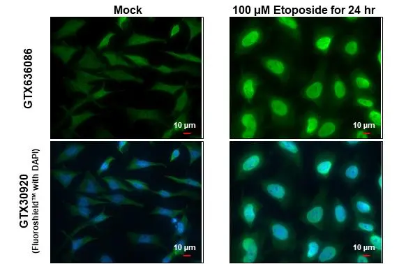
ATM (phospho Ser1981) antibody [HL1062] detects ATM (phospho Ser1981) protein at nucleus by immunofluorescent analysis. Sample: Mock and treated HeLa cells were fixed in 4% paraformaldehyde at RT for 15 min. Green: ATM (phospho Ser1981) stained by ATM (phospho Ser1981) antibody [HL1062] (GTX636086) diluted at 1:500. Blue: Fluoroshield with DAPI (GTX30920).
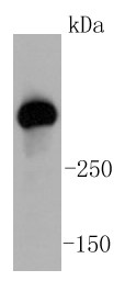
![Untreated (–) and treated (+) 293T whole cell extracts (60 μg) were separated by 5% SDS-PAGE, and the membrane was blotted with ATM (phospho Ser1981) antibody [HL1062] (GTX636086) diluted at 1:1000. The HRP-conjugated anti-rabbit IgG antibody (GTX213110-01) was used to detect the primary antibody.](https://www.genetex.com/upload/website/prouct_img/normal/GTX636086/GTX636086_44333_20210604_WB_treatment_UVC_w_23061202_695.webp "Untreated (–) and treated (+) 293T whole cell extracts (60 μg) were separated by 5% SDS-PAGE, and the membrane was blotted with ATM (phospho Ser1981) antibody [HL1062] (GTX636086) diluted at 1:1000. The HRP-conjugated anti-rabbit IgG antibody (GTX213110-01) was used to detect the primary antibody.")
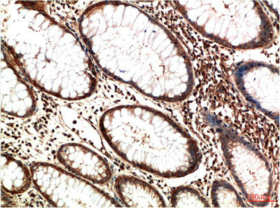
![ATM (phospho Ser1981) antibody [HL1062] detects ATM (phospho Ser1981) protein at nucleus by immunohistochemical analysis. Sample: Paraffin-embedded human cervical carcinoma. ATM (phospho Ser1981) stained by ATM (phospho Ser1981) antibody [HL1062] (GTX636086) diluted at 1:100. Antigen Retrieval: Citrate buffer, pH 6.0, 15 min](https://www.genetex.com/upload/website/prouct_img/normal/GTX636086/GTX636086_44333_20210702_IHC-P_w_23061202_647.webp "ATM (phospho Ser1981) antibody [HL1062] detects ATM (phospho Ser1981) protein at nucleus by immunohistochemical analysis. Sample: Paraffin-embedded human cervical carcinoma. ATM (phospho Ser1981) stained by ATM (phospho Ser1981) antibody [HL1062] (GTX636086) diluted at 1:100. Antigen Retrieval: Citrate buffer, pH 6.0, 15 min")
![Untreated (–) and treated (+) 293T whole cell extracts (60 μg) were separated by 5% SDS-PAGE, and the membrane was blotted with ATM (phospho Ser1981) antibody [HL1062] (GTX636086) diluted at 1:1000. The HRP-conjugated anti-rabbit IgG antibody (GTX213110-01) was used to detect the primary antibody, and the signal was developed with Trident ECL plus-Enhanced.](https://www.genetex.com/upload/website/prouct_img/normal/GTX636086/GTX636086_44333_20220128_WB_treatment_UVC_peptideblocking_w_23061202_850.webp "Untreated (–) and treated (+) 293T whole cell extracts (60 μg) were separated by 5% SDS-PAGE, and the membrane was blotted with ATM (phospho Ser1981) antibody [HL1062] (GTX636086) diluted at 1:1000. The HRP-conjugated anti-rabbit IgG antibody (GTX213110-01) was used to detect the primary antibody, and the signal was developed with Trident ECL plus-Enhanced.")
![Untreated (–) and treated (+) 293T whole cell extracts were separated by 5% SDS-PAGE, and the membrane was blotted with ATM (phospho Ser1981) antibody [HL1062] (GTX636086) diluted at 1:1000. The HRP-conjugated anti-rabbit IgG antibody (GTX213110-01) was used to detect the primary antibody.](https://www.genetex.com/upload/website/prouct_img/normal/GTX636086/GTX636086_45397_20240503_WB_treatment_UVC_24101600_996.webp "Untreated (–) and treated (+) 293T whole cell extracts were separated by 5% SDS-PAGE, and the membrane was blotted with ATM (phospho Ser1981) antibody [HL1062] (GTX636086) diluted at 1:1000. The HRP-conjugated anti-rabbit IgG antibody (GTX213110-01) was used to detect the primary antibody.")
![Untreated (–) and treated (+) 293T whole cell extracts (60 μg) were separated by 5% SDS-PAGE, and the membrane was blotted with ATM (phospho Ser1981) antibody [HL1062] (GTX636086) diluted at 1:1000. The HRP-conjugated anti-rabbit IgG antibody (GTX213110-01) was used to detect the primary antibody.](https://www.genetex.com/upload/website/prouct_img/normal/GTX636086/GTX636086_45271_20240105_WB_treatment_UVC_24101600_953.webp "Untreated (–) and treated (+) 293T whole cell extracts (60 μg) were separated by 5% SDS-PAGE, and the membrane was blotted with ATM (phospho Ser1981) antibody [HL1062] (GTX636086) diluted at 1:1000. The HRP-conjugated anti-rabbit IgG antibody (GTX213110-01) was used to detect the primary antibody.")
ATM (phospho Ser1981) antibody [HL1062] detects ATM (phospho Ser1981) protein at nucleus by immunofluorescent analysis. Sample: Mock and treated HeLa cells were fixed in 4% paraformaldehyde at RT for 15 min. Green: ATM (phospho Ser1981) stained by ATM (phospho Ser1981) antibody [HL1062] (GTX636086) diluted at 1:500. Blue: Fluoroshield with DAPI (GTX30920).
ATM (phospho Ser1981) antibody [HL1062]
GTX636086
ApplicationsImmunoFluorescence, Western Blot, ImmunoCytoChemistry, ImmunoHistoChemistry, ImmunoHistoChemistry Paraffin
Product group Antibodies
ReactivityHuman
TargetATM
Overview
- SupplierGeneTex
- Product NameATM (phospho Ser1981) antibody [HL1062]
- Delivery Days Customer9
- Application Supplier NoteWB: 1:500-1:3000. *Optimal dilutions/concentrations should be determined by the researcher.Not tested in other applications.
- ApplicationsImmunoFluorescence, Western Blot, ImmunoCytoChemistry, ImmunoHistoChemistry, ImmunoHistoChemistry Paraffin
- CertificationResearch Use Only
- ClonalityMonoclonal
- Clone IDHL1062
- Concentration1 mg/ml
- ConjugateUnconjugated
- Gene ID472
- Target nameATM
- Target descriptionATM serine/threonine kinase
- Target synonymsAT1, ATA, ATC, ATD, ATDC, ATE, TEL1, TELO1, serine-protein kinase ATM, A-T mutated, AT mutated, TEL1, telomere maintenance 1, homolog, ataxia telangiectasia mutated, serine/threonine kinase ATM
- HostRabbit
- IsotypeIgG
- Protein IDQ13315
- Protein NameSerine-protein kinase ATM
- Scientific DescriptionThe protein encoded by this gene belongs to the PI3/PI4-kinase family. This protein is an important cell cycle checkpoint kinase that phosphorylates; thus, it functions as a regulator of a wide variety of downstream proteins, including tumor suppressor proteins p53 and BRCA1, checkpoint kinase CHK2, checkpoint proteins RAD17 and RAD9, and DNA repair protein NBS1. This protein and the closely related kinase ATR are thought to be master controllers of cell cycle checkpoint signaling pathways that are required for cell response to DNA damage and for genome stability. Mutations in this gene are associated with ataxia telangiectasia, an autosomal recessive disorder. [provided by RefSeq, Aug 2010]
- ReactivityHuman
- Storage Instruction-20°C or -80°C,2°C to 8°C
- UNSPSC41116161
Datasheet
Related products

Product group Antibodies
Anti-ATM AntibodyA99484
ApplicationsELISA, ImmunoHistoChemistry
ReactivityHuman, Mouse
- SizePrice
Product group Antibodies
Anti-ATM Antibody144-05908
ApplicationsWestern Blot
ReactivityHuman
TargetATM
- SizePrice
Product group Antibodies
ATM Recombinant AntibodyBSM-52360R
ApplicationsImmunoFluorescence, Western Blot, ImmunoCytoChemistry, ImmunoHistoChemistry, ImmunoHistoChemistry Frozen, ImmunoHistoChemistry Paraffin
ReactivityHuman
TargetATM
- SizePrice
Product group Antibodies
ATM Monoclonal AntibodyCSB-MA341522
ApplicationsELISA, ImmunoHistoChemistry
ReactivityHuman, Mouse, Rat
TargetATM
- SizePrice
Product group Antibodies
ApplicationsImmunoFluorescence, Western Blot, ImmunoHistoChemistry
TargetATM
- SizePrice
Product group Antibodies
ATM (phospho Ser1981) antibodyGTX132146
ApplicationsImmunoFluorescence, Western Blot, ImmunoCytoChemistry, ImmunoHistoChemistry, ImmunoHistoChemistry Paraffin
ReactivityHuman, Mouse
TargetATM
- SizePrice
Product group Antibodies
ATM antibodyGTX132147
ApplicationsImmunoFluorescence, Western Blot, ImmunoCytoChemistry
ReactivityHuman
TargetATM
- SizePrice
![ICC/IF analysis of 293 untreated cells (left panel) or stimulated cells with 100 mJ UV (right panel) using GTX15666 ATM (phospho Ser1981) antibody [10H11]. Fixation : Formalin Permeabilization : 0.1% Triton X-100 in TBS for 5-10 minutes](https://www.genetex.com/upload/website/prouct_img/normal/GTX15666/GTX15666_280_ICC-IF_w_23060620_691.webp)
Product group Antibodies
ApplicationsImmunoFluorescence, ImmunoPrecipitation, Western Blot, ImmunoCytoChemistry
ReactivityHuman, Mouse
TargetATM
- SizePrice
Product group Antibodies
ATM antibodyGTX22631
ApplicationsImmunoPrecipitation, Western Blot
ReactivityHuman, Mouse
TargetATM
- SizePrice