

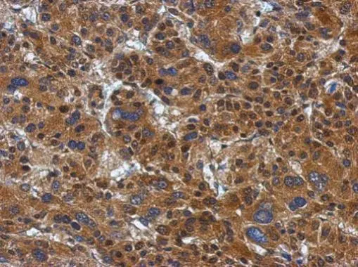
Immunohistochemical analysis of paraffin-embedded Hepatoma, using ATP citrate lyase(GTX112387) antibody at 1:500 dilution.
Antigen Retrieval: Trilogy? (EDTA based, pH 8.0) buffer, 15min
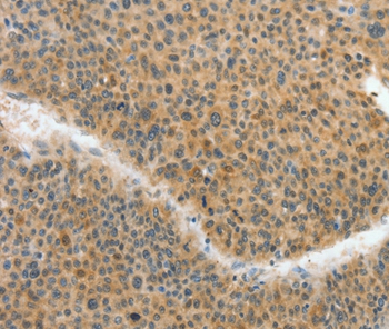
![ATP citrate lyase antibody [N1N2], N-term detects ATP citrate lyase protein at cytoplasm by immunohistochemical analysis. Sample: Paraffin-embedded mouse pancreas. ATP citrate lyase stained by ATP citrate lyase antibody [N1N2], N-term (GTX112387) diluted at 1:500. Antigen Retrieval: Citrate buffer, pH 6.0, 15 min](https://www.genetex.com/upload/website/prouct_img/normal/GTX112387/GTX112387_44000_20210625_IHC-P_M_w_23060500_241.webp "ATP citrate lyase antibody [N1N2], N-term detects ATP citrate lyase protein at cytoplasm by immunohistochemical analysis. Sample: Paraffin-embedded mouse pancreas. ATP citrate lyase stained by ATP citrate lyase antibody [N1N2], N-term (GTX112387) diluted at 1:500. Antigen Retrieval: Citrate buffer, pH 6.0, 15 min")
![ATP citrate lyase antibody [N1N2], N-term detects ATP citrate lyase protein at cytoplasm by immunofluorescent analysis. Sample: HeLa cells were fixed in ice-cold MeOH for 5 min. Green: ATP citrate lyase protein stained by ATP citrate lyase antibody [N1N2], N-term (GTX112387) diluted at 1:500. Blue: Hoechst 33342 staining.](https://www.genetex.com/upload/website/prouct_img/normal/GTX112387/GTX112387_40667_20171012_IFA_w_23060500_779.webp "ATP citrate lyase antibody [N1N2], N-term detects ATP citrate lyase protein at cytoplasm by immunofluorescent analysis. Sample: HeLa cells were fixed in ice-cold MeOH for 5 min. Green: ATP citrate lyase protein stained by ATP citrate lyase antibody [N1N2], N-term (GTX112387) diluted at 1:500. Blue: Hoechst 33342 staining.")
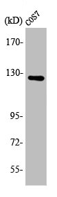
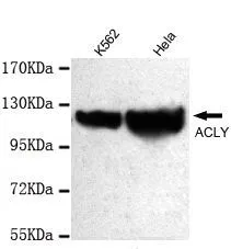
![Various tissue extracts (50 μg) were separated by 5% SDS-PAGE, and the membrane was blotted with ATP citrate lyase antibody [N1N2], N-term (GTX112387) diluted at 1:5000. The HRP-conjugated anti-rabbit IgG antibody (GTX213110-01) was used to detect the primary antibody.](https://www.genetex.com/upload/website/prouct_img/normal/GTX112387/GTX112387_40667_20180330_WB_M_R_w_23060500_849.webp "Various tissue extracts (50 μg) were separated by 5% SDS-PAGE, and the membrane was blotted with ATP citrate lyase antibody [N1N2], N-term (GTX112387) diluted at 1:5000. The HRP-conjugated anti-rabbit IgG antibody (GTX213110-01) was used to detect the primary antibody.")
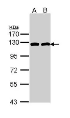
![Various whole cell extracts (30 μg) were separated by 5% SDS-PAGE, and the membrane was blotted with ATP citrate lyase antibody [N1N2], N-term (GTX112387) diluted at 1:5000. The HRP-conjugated anti-rabbit IgG antibody (GTX213110-01) was used to detect the primary antibody. Corresponding RNA expression data for the same cell lines are based on Human Protein Atlas program.](https://www.genetex.com/upload/website/prouct_img/normal/GTX112387/GTX112387_45105_20230714_WB_TPM_watermark_24072519_808.webp "Various whole cell extracts (30 μg) were separated by 5% SDS-PAGE, and the membrane was blotted with ATP citrate lyase antibody [N1N2], N-term (GTX112387) diluted at 1:5000. The HRP-conjugated anti-rabbit IgG antibody (GTX213110-01) was used to detect the primary antibody. Corresponding RNA expression data for the same cell lines are based on Human Protein Atlas program.")
Immunohistochemical analysis of paraffin-embedded Hepatoma, using ATP citrate lyase(GTX112387) antibody at 1:500 dilution.
Antigen Retrieval: Trilogy? (EDTA based, pH 8.0) buffer, 15min
ATP citrate lyase antibody [N1N2], N-term
GTX112387
ApplicationsImmunoFluorescence, Western Blot, ImmunoCytoChemistry, ImmunoHistoChemistry, ImmunoHistoChemistry Paraffin
Product group Antibodies
ReactivityHuman, Mouse, Rat
TargetACLY
Overview
- SupplierGeneTex
- Product NameATP citrate lyase antibody [N1N2], N-term
- Delivery Days Customer9
- Application Supplier NoteWB: 1:1000-1:10000. ICC/IF: 1:100-1:1000. IHC-P: 1:100-1:1000. *Optimal dilutions/concentrations should be determined by the researcher.Not tested in other applications.
- ApplicationsImmunoFluorescence, Western Blot, ImmunoCytoChemistry, ImmunoHistoChemistry, ImmunoHistoChemistry Paraffin
- CertificationResearch Use Only
- ClonalityPolyclonal
- Concentration0.54 mg/ml
- ConjugateUnconjugated
- Gene ID47
- Target nameACLY
- Target descriptionATP citrate lyase
- Target synonymsACL, ATPCL, CLATP, ATP-citrate synthase, ATP-citrate (pro-S-)-lyase, citrate cleavage enzyme
- HostRabbit
- IsotypeIgG
- Protein IDP53396
- Protein NameATP-citrate synthase
- Scientific DescriptionATP citrate lyase is the primary enzyme responsible for the synthesis of cytosolic acetyl-CoA in many tissues. The enzyme is a tetramer (relative molecular weight approximately 440,000) of apparently identical subunits. It catalyzes the formation of acetyl-CoA and oxaloacetate from citrate and CoA with a concomitant hydrolysis of ATP to ADP and phosphate. The product, acetyl-CoA, serves several important biosynthetic pathways, including lipogenesis and cholesterogenesis. In nervous tissue, ATP citrate-lyase may be involved in the biosynthesis of acetylcholine. Two transcript variants encoding distinct isoforms have been identified for this gene. [provided by RefSeq]
- ReactivityHuman, Mouse, Rat
- Storage Instruction-20°C or -80°C,2°C to 8°C
- UNSPSC41116161
Datasheet
Related products
Product group Antibodies
Anti-ACLY AntibodyA46777
ApplicationsImmunoHistoChemistry
ReactivityHuman
- SizePrice
Product group Antibodies
Anti-ACLY Antibody144-60960
ApplicationsWestern Blot
ReactivityHuman, Mouse, Rat
TargetACLY
- SizePrice

Product group Antibodies
ACLY Recombinant AntibodyBSM-52458R
ApplicationsFlow Cytometry, ImmunoFluorescence, Western Blot, ImmunoCytoChemistry, ImmunoHistoChemistry, ImmunoHistoChemistry Frozen, ImmunoHistoChemistry Paraffin
ReactivityHuman, Mouse, Rat
TargetACLY
- SizePrice
Product group Antibodies
ACLY AntibodyCSB-PA000957
ApplicationsImmunoFluorescence, Western Blot, ELISA
ReactivityHuman, Monkey, Mouse, Rat
TargetACLY
- SizePrice
Product group Antibodies
Acly Polyclonal AntibodyCAC08207
ApplicationsImmunoFluorescence, Western Blot, ELISA, ImmunoHistoChemistry
TargetACLY
- SizePrice
Product group Antibodies

ACLY / ATP Citrate Lyase AntibodyLS-C401433
ApplicationsELISA, ImmunoHistoChemistry
ReactivityHuman, Mouse, Rat
TargetACLY
- SizePrice
![IHC-P analysis of colon adenocarcinoma tissue using GTX84967 ATP citrate lyase antibody [3G8]. Antigen retrieval : Heat-induced epitope retrieval by 10mM citrate buffer, pH6.0, 100oC for 10min. Dilution : 1:50](https://www.genetex.com/upload/website/prouct_img/normal/GTX84967/GTX84967_3302_IHC-P_w_23061420_547.webp)
Product group Antibodies
ATP citrate lyase antibody [3G8]GTX84967
ApplicationsImmunoFluorescence, Western Blot, ImmunoCytoChemistry, ImmunoHistoChemistry, ImmunoHistoChemistry Paraffin
ReactivityHuman, Monkey, Mouse
TargetACLY
- SizePrice

Product group Antibodies
Anti-ACLY AntibodyHPA022434
ApplicationsWestern Blot, ImmunoCytoChemistry, ImmunoHistoChemistry
ReactivityHuman, Mouse, Rat
TargetACLY
- SizePrice
Product group Antibodies
ApplicationsImmunoFluorescence, Western Blot, ImmunoCytoChemistry
ReactivityHuman, Mouse, Rat
TargetACLY
- SizePrice
Product group Antibodies
ApplicationsFlow Cytometry, ImmunoFluorescence, Western Blot, ImmunoCytoChemistry
ReactivityHuman
TargetACLY
- SizePrice