

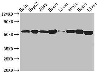
Western Blot Positive WB detected in: Hela whole cell lysate, HepG2 whole cell lysate, A549 whole cell lysate, Mouse heart tissue, Mouse liver tissue, Mouse brain tissue, Rat heart tissue, Rat liver tissue All lanes: ATP5B antibody at 2.7microg/ml Secondary Goat polyclonal to rabbit IgG at 1/50000 dilution Predicted band size: 57 kDa Observed band size: 57 kDa


Western Blot Positive WB detected in: Hela whole cell lysate, HepG2 whole cell lysate, A549 whole cell lysate, Mouse heart tissue, Mouse liver tissue, Mouse brain tissue, Rat heart tissue, Rat liver tissue All lanes: ATP5B antibody at 2.7microg/ml Secondary Goat polyclonal to rabbit IgG at 1/50000 dilution Predicted band size: 57 kDa Observed band size: 57 kDa
ATP5B Antibody
CSB-PA002350EA01HU
ApplicationsWestern Blot, ELISA, ImmunoHistoChemistry
Product group Antibodies
ReactivityHuman, Mouse, Rat
TargetATP5F1B
Overview
- SupplierCusabio
- Product NameATP5B Antibody
- Delivery Days Customer20
- ApplicationsWestern Blot, ELISA, ImmunoHistoChemistry
- CertificationResearch Use Only
- ClonalityPolyclonal
- ConjugateUnconjugated
- Gene ID506
- Target nameATP5F1B
- Target descriptionATP synthase F1 subunit beta
- Target synonymsATP5B, ATPMB, ATPSB, HEL-S-271, HUMOP2, ATP synthase F(1) complex subunit beta, mitochondrial, ATP synthase subunit beta, mitochondrial, ATP synthase, H+ transporting, mitochondrial F1 complex, beta polypeptide, epididymis secretory protein Li 271, mitochondrial ATP synthase beta subunit, mitochondrial ATP synthetase, beta subunit
- HostRabbit
- IsotypeIgG
- Protein IDP06576
- Protein NameATP synthase F(1) complex subunit beta, mitochondrial
- Scientific DescriptionMitochondrial membrane ATP synthase (F(1)F(0) ATP synthase or Complex V) produces ATP from ADP in the presence of a proton gradient across the membrane which is generated by electron transport complexes of the respiratory chain. F-type ATPases consist of two structural domains, F(1) - containing the extramembraneous catalytic core, and F(0) - containing the membrane proton channel, linked together by a central stalk and a peripheral stalk. During catalysis, ATP synthesis in the catalytic domain of F(1) is coupled via a rotary mechanism of the central stalk subunits to proton translocation. Subunits alpha and beta form the catalytic core in F(1). Rotation of the central stalk against the surrounding alpha(3)beta(3) subunits leads to hydrolysis of ATP in three separate catalytic sites on the beta subunits.
- ReactivityHuman, Mouse, Rat
- Storage Instruction-20°C or -80°C
- UNSPSC41116161
Related products

Product group Antibodies
Anti-ATP5B AntibodyA31039
ApplicationsWestern Blot, ImmunoHistoChemistry
ReactivityHuman, Mouse, Rat
- SizePrice
Product group Antibodies
Anti-ATP5B Antibody144-05769
ApplicationsImmunoFluorescence, Western Blot, ImmunoHistoChemistry
ReactivityHuman, Mouse, Rat
TargetATP5F1B
- SizePrice

Product group Antibodies
Anti-ATP5F1B Antibody Picoband(r)A32270-3-CARRIER-FREE
ApplicationsFlow Cytometry, ImmunoFluorescence, Western Blot, ELISA, ImmunoCytoChemistry, ImmunoHistoChemistry
ReactivityHuman, Mouse, Rat
TargetATP5F1B
- SizePrice
Product group Antibodies
ApplicationsFlow Cytometry, Western Blot, ImmunoCytoChemistry
ReactivityHuman, Mouse, Rat
TargetATP5F1B
- SizePrice
Product group Antibodies

Goat anti-ATP5B (aa15162)EB12459
ApplicationsWestern Blot, ELISA
ReactivityBovine, Canine, Human, Mouse, Porcine, Rat
TargetATP5F1B
- SizePrice
Product group Antibodies
ATP5B Polyclonal AntibodyCAC14816
ApplicationsWestern Blot, ELISA, ImmunoHistoChemistry
ReactivityMouse, Rat
TargetATP5F1B
- SizePrice
Product group Antibodies
ATP5B antibodyGTX132925
ApplicationsImmunoFluorescence, Western Blot, ImmunoCytoChemistry
ReactivityHuman
TargetATP5F1B
- SizePrice
Product group Antibodies

ATP5B / ATP Synthase Beta AntibodyLS-C482498
ApplicationsWestern Blot, ImmunoHistoChemistry, ImmunoHistoChemistry Paraffin
ReactivityHuman, Mouse, Rat
TargetATP5F1B
- SizePrice

Product group Antibodies
Anti-ATP5B AntibodyHPA001520
ApplicationsWestern Blot, ImmunoCytoChemistry, ImmunoHistoChemistry
ReactivityHuman, Mouse, Rat
TargetATP5F1B
- SizePrice