

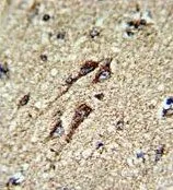
IHC-P analysis of human brain tissue using GTX81939 ATP5B antibody, Internal.


 either nontransfected (Lane 1) or transiently transfected with the ATP5B (Lane 2) using GTX81939 ATP5B antibody, Internal.")

IHC-P analysis of human brain tissue using GTX81939 ATP5B antibody, Internal.
ATP5B antibody, Internal
GTX81939
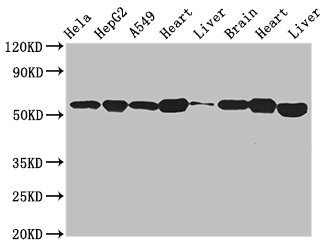
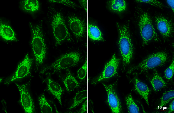
ApplicationsFlow Cytometry, ImmunoFluorescence, Western Blot, ImmunoCytoChemistry, ImmunoHistoChemistry, ImmunoHistoChemistry Paraffin
Product group Antibodies
ReactivityHuman
TargetATP5F1B
Overview
- SupplierGeneTex
- Product NameATP5B antibody, Internal
- Delivery Days Customer9
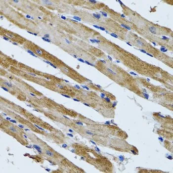
- Application Supplier NoteWB: 1:1000. ICC/IF: 1:25. IHC-P: 1:50-1:100. FCM: 1:10-1:50. *Optimal dilutions/concentrations should be determined by the researcher.Not tested in other applications.
- ApplicationsFlow Cytometry, ImmunoFluorescence, Western Blot, ImmunoCytoChemistry, ImmunoHistoChemistry, ImmunoHistoChemistry Paraffin
- CertificationResearch Use Only
- ClonalityPolyclonal
- ConjugateUnconjugated
- Gene ID506
- Target nameATP5F1B
- Target descriptionATP synthase F1 subunit beta
- Target synonymsATP5B, ATPMB, ATPSB, HEL-S-271, HUMOP2, ATP synthase F(1) complex subunit beta, mitochondrial, ATP synthase subunit beta, mitochondrial, ATP synthase, H+ transporting, mitochondrial F1 complex, beta polypeptide, epididymis secretory protein Li 271, mitochondrial ATP synthase beta subunit, mitochondrial ATP synthetase, beta subunit
- HostRabbit
- IsotypeIgG
- Protein IDP06576
- Protein NameATP synthase F(1) complex subunit beta, mitochondrial
- Scientific DescriptionThis gene encodes a subunit of mitochondrial ATP synthase. Mitochondrial ATP synthase catalyzes ATP synthesis, utilizing an electrochemical gradient of protons across the inner membrane during oxidative phosphorylation. ATP synthase is composed of two linked multi-subunit complexes: the soluble catalytic core, F1, and the membrane-spanning component, Fo, comprising the proton channel. The catalytic portion of mitochondrial ATP synthase consists of 5 different subunits (alpha, beta, gamma, delta, and epsilon) assembled with a stoichiometry of 3 alpha, 3 beta, and a single representative of the other 3. The proton channel consists of three main subunits (a, b, c). This gene encodes the beta subunit of the catalytic core. [provided by RefSeq, Jul 2008]
- ReactivityHuman
- Storage Instruction-20°C or -80°C,2°C to 8°C
- UNSPSC41116161
Datasheet
Related products

Product group Antibodies
Anti-ATP5B AntibodyA31039
ApplicationsWestern Blot, ImmunoHistoChemistry
ReactivityHuman, Mouse, Rat
- SizePrice
Product group Antibodies
Anti-ATP5B Antibody144-05769
ApplicationsImmunoFluorescence, Western Blot, ImmunoHistoChemistry
ReactivityHuman, Mouse, Rat
TargetATP5F1B
- SizePrice

Product group Antibodies
Anti-ATP5F1B Antibody Picoband(r)A32270-3-CARRIER-FREE
ApplicationsFlow Cytometry, ImmunoFluorescence, Western Blot, ELISA, ImmunoCytoChemistry, ImmunoHistoChemistry
ReactivityHuman, Mouse, Rat
TargetATP5F1B
- SizePrice
Product group Antibodies
ApplicationsFlow Cytometry, Western Blot, ImmunoCytoChemistry
ReactivityHuman, Mouse, Rat
TargetATP5F1B
- SizePrice
Product group Antibodies
ATP5B AntibodyCSB-PA002350EA01HU
ApplicationsWestern Blot, ELISA, ImmunoHistoChemistry
ReactivityHuman, Mouse, Rat
TargetATP5F1B
- SizePrice
Product group Antibodies

Goat anti-ATP5B (aa15162)EB12459
ApplicationsWestern Blot, ELISA
ReactivityBovine, Canine, Human, Mouse, Porcine, Rat
TargetATP5F1B
- SizePrice
Product group Antibodies
ATP5B Polyclonal AntibodyCAC14816
ApplicationsWestern Blot, ELISA, ImmunoHistoChemistry
ReactivityMouse, Rat
TargetATP5F1B
- SizePrice
Product group Antibodies
ATP5B antibodyGTX132925
ApplicationsImmunoFluorescence, Western Blot, ImmunoCytoChemistry
ReactivityHuman
TargetATP5F1B
- SizePrice
Product group Antibodies
ATP5B antibodyGTX30075
ApplicationsImmunoFluorescence, Western Blot, ImmunoCytoChemistry, ImmunoHistoChemistry, ImmunoHistoChemistry Paraffin
ReactivityHuman, Mouse, Rat
TargetATP5F1B
- SizePrice
Product group Antibodies

ATP5B / ATP Synthase Beta AntibodyLS-C482498
ApplicationsWestern Blot, ImmunoHistoChemistry, ImmunoHistoChemistry Paraffin
ReactivityHuman, Mouse, Rat
TargetATP5F1B
- SizePrice