

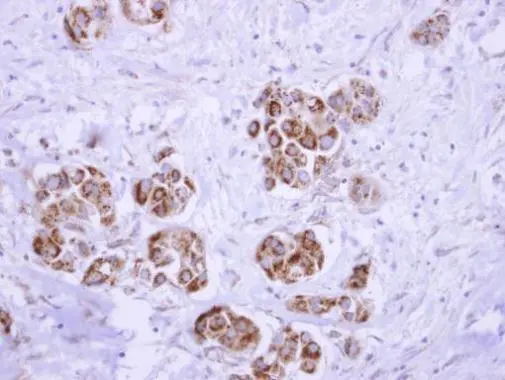
ATP synthase delta antibody detects ATP5D protein at mitochondria on human breast cancer by immunohistochemical analysis. Sample: Paraffin-embedded breast cancer. ATP synthase delta antibody (GTX101503) dilution: 1:250.
Antigen Retrieval: Trilogy? (EDTA based, pH 8.0) buffer, 15min
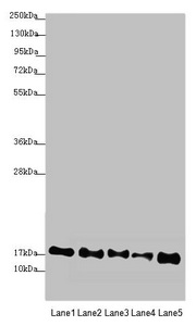
 A: Mouse brain 12% SDS PAGE GTX101503 diluted at 1:1000")
 were separated by SDS-PAGE, and the membrane was blotted with ATP5D antibody (GTX101503) diluted at 1:2000. The HRP-conjugated anti-rabbit IgG antibody (GTX213110-01) was used to detect the primary antibody.")
ATP synthase delta antibody detects ATP5D protein at mitochondria on human breast cancer by immunohistochemical analysis. Sample: Paraffin-embedded breast cancer. ATP synthase delta antibody (GTX101503) dilution: 1:250.
Antigen Retrieval: Trilogy? (EDTA based, pH 8.0) buffer, 15min
ATP5D antibody
GTX101503
ApplicationsWestern Blot, ImmunoHistoChemistry, ImmunoHistoChemistry Paraffin
Product group Antibodies
ReactivityHuman, Mouse
TargetATP5F1D
Overview
- SupplierGeneTex
- Product NameATP5D antibody
- Delivery Days Customer9
- Application Supplier NoteWB: 1:500-1:3000. IHC-P: 1:100-1:1000. *Optimal dilutions/concentrations should be determined by the researcher.Not tested in other applications.
- ApplicationsWestern Blot, ImmunoHistoChemistry, ImmunoHistoChemistry Paraffin
- CertificationResearch Use Only
- ClonalityPolyclonal
- Concentration1 mg/ml
- ConjugateUnconjugated
- Gene ID513
- Target nameATP5F1D
- Target descriptionATP synthase F1 subunit delta
- Target synonymsATP5D, MC5DN5, ATP synthase F(1) complex subunit delta, mitochondrial, ATP synthase subunit delta, mitochondrial, ATP synthase, H+ transporting, mitochondrial F1 complex, delta subunit, F-ATPase delta subunit, mitochondrial ATP synthase complex delta-subunit precusor, mitochondrial ATP synthase, delta subunit
- HostRabbit
- IsotypeIgG
- Protein IDP30049
- Protein NameATP synthase F(1) complex subunit delta, mitochondrial
- Scientific DescriptionThis gene encodes a subunit of mitochondrial ATP synthase. Mitochondrial ATP synthase catalyzes ATP synthesis, utilizing an electrochemical gradient of protons across the inner membrane during oxidative phosphorylation. ATP synthase is composed of two linked multi-subunit complexes: the soluble catalytic core, F1, and the membrane-spanning component, Fo, comprising the proton channel. The catalytic portion of mitochondrial ATP synthase consists of 5 different subunits (alpha, beta, gamma, delta, and epsilon) assembled with a stoichiometry of 3 alpha, 3 beta, and a single representative of the other 3. The proton channel consists of three main subunits (a, b, c). This gene encodes the delta subunit of the catalytic core. Alternatively spliced transcript variants encoding the same isoform have been identified. [provided by RefSeq]
- ReactivityHuman, Mouse
- Storage Instruction-20°C or -80°C,2°C to 8°C
- UNSPSC41116161
Datasheet
Related products
Product group Antibodies
Anti-ATP5D Antibody144-09929
ApplicationsWestern Blot
ReactivityHuman, Mouse, Rat
TargetATP5F1D
- SizePrice

Product group Antibodies
Anti-ATP5F1D Antibody Picoband(r)A32272-2-CARRIER-FREE
ApplicationsImmunoFluorescence, Western Blot, ELISA, ImmunoCytoChemistry, ImmunoHistoChemistry
ReactivityHuman, Mouse, Rat
TargetATP5F1D
- SizePrice
Product group Antibodies
Anti-ATP5D AntibodyA95933
ApplicationsImmunoFluorescence, ELISA, ImmunoHistoChemistry
ReactivityHuman, Mouse, Rat
- SizePrice
Product group Antibodies
Atp5F1D Polyclonal AntibodyCAC07345
ApplicationsWestern Blot, ELISA, ImmunoHistoChemistry
TargetATP5F1D
- SizePrice
Product group Antibodies
ATP5F1D AntibodyCSB-PA002355ESR1HU
ApplicationsWestern Blot, ELISA
ReactivityHuman, Mouse
TargetATP5F1D
- SizePrice
Product group Antibodies
ATP5D antibodyGTX66588
ApplicationsImmunoFluorescence, Western Blot, ImmunoCytoChemistry
ReactivityHuman, Mouse, Rat
TargetATP5F1D
- SizePrice

Product group Antibodies
Anti-ATP5D AntibodyHPA002865
ApplicationsWestern Blot, ImmunoHistoChemistry
ReactivityHuman, Mouse, Rat
TargetATP5F1D
- SizePrice
Product group Antibodies

ATP5D AntibodyLS-C830896
ApplicationsELISA, ImmunoHistoChemistry
ReactivityHuman, Mouse, Rat
TargetATP5F1D
- SizePrice
Product group Antibodies
Anti-ATP5D AntibodyCAB9929
ApplicationsImmunoFluorescence, Western Blot, ELISA, ImmunoCytoChemistry
ReactivityHuman
- SizePrice