

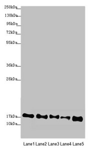
Western blot All lanes: ATP5F1D antibody at 2.44microg/ml Lane 1: Mouse heart tissue Lane 2: Raji whole cell lysate Lane 3: NIH/3T3 whole cell lysate Lane 4: A549 whole cell lysate Lane 5: HepG2 whole cell lysate Secondary Goat polyclonal to rabbit IgG at 1/10000 dilution Predicted band size: 18 kDa Observed band size: 18 kDa
Western blot All lanes: ATP5F1D antibody at 2.44microg/ml Lane 1: Mouse heart tissue Lane 2: Raji whole cell lysate Lane 3: NIH/3T3 whole cell lysate Lane 4: A549 whole cell lysate Lane 5: HepG2 whole cell lysate Secondary Goat polyclonal to rabbit IgG at 1/10000 dilution Predicted band size: 18 kDa Observed band size: 18 kDa
ATP5F1D Antibody
CSB-PA002355ESR1HU
ApplicationsWestern Blot, ELISA
Product group Antibodies
ReactivityHuman, Mouse
TargetATP5F1D
Overview
- SupplierCusabio
- Product NameATP5F1D Antibody
- Delivery Days Customer20
- ApplicationsWestern Blot, ELISA
- CertificationResearch Use Only
- ClonalityPolyclonal
- ConjugateUnconjugated
- Gene ID513
- Target nameATP5F1D
- Target descriptionATP synthase F1 subunit delta
- Target synonymsATP5D, MC5DN5, ATP synthase F(1) complex subunit delta, mitochondrial, ATP synthase subunit delta, mitochondrial, ATP synthase, H+ transporting, mitochondrial F1 complex, delta subunit, F-ATPase delta subunit, mitochondrial ATP synthase complex delta-subunit precusor, mitochondrial ATP synthase, delta subunit
- HostRabbit
- IsotypeIgG
- Protein IDP30049
- Protein NameATP synthase F(1) complex subunit delta, mitochondrial
- Scientific DescriptionMitochondrial membrane ATP synthase (F(1)F(0) ATP synthase or Complex V) produces ATP from ADP in the presence of a proton gradient across the membrane which is generated by electron transport complexes of the respiratory chain. F-type ATPases consist of two structural domains, F(1) - containing the extramembraneous catalytic core, and F(0) - containing the membrane proton channel, linked together by a central stalk and a peripheral stalk. During catalysis, ATP turnover in the catalytic domain of F(1) is coupled via a rotary mechanism of the central stalk subunits to proton translocation. Part of the complex F(1) domain and of the central stalk which is part of the complex rotary element. Rotation of the central stalk against the surrounding alpha(3)beta(3) subunits leads to hydrolysis of ATP in three separate catalytic sites on the beta subunits.
- ReactivityHuman, Mouse
- Storage Instruction-20°C or -80°C
- UNSPSC41116161
Related products
Product group Antibodies
Anti-ATP5D AntibodyA95933
ApplicationsImmunoFluorescence, ELISA, ImmunoHistoChemistry
ReactivityHuman, Mouse, Rat
- SizePrice
Product group Antibodies
Anti-ATP5D Antibody144-09929
ApplicationsWestern Blot
ReactivityHuman, Mouse, Rat
TargetATP5F1D
- SizePrice

Product group Antibodies
Anti-ATP5F1D Antibody Picoband(r)A32272-2-CARRIER-FREE
ApplicationsImmunoFluorescence, Western Blot, ELISA, ImmunoCytoChemistry, ImmunoHistoChemistry
ReactivityHuman, Mouse, Rat
TargetATP5F1D
- SizePrice
Product group Antibodies

ATP5D AntibodyLS-C830896
ApplicationsELISA, ImmunoHistoChemistry
ReactivityHuman, Mouse, Rat
TargetATP5F1D
- SizePrice
Product group Antibodies
Atp5F1D Polyclonal AntibodyCAC07345
ApplicationsWestern Blot, ELISA, ImmunoHistoChemistry
TargetATP5F1D
- SizePrice
Product group Antibodies
ATP5D antibodyGTX101503
ApplicationsWestern Blot, ImmunoHistoChemistry, ImmunoHistoChemistry Paraffin
ReactivityHuman, Mouse
TargetATP5F1D
- SizePrice

Product group Antibodies
Anti-ATP5D AntibodyHPA002865
ApplicationsWestern Blot, ImmunoHistoChemistry
ReactivityHuman, Mouse, Rat
TargetATP5F1D
- SizePrice
Product group Antibodies
Anti-ATP5D AntibodyCAB9929
ApplicationsImmunoFluorescence, Western Blot, ELISA, ImmunoCytoChemistry
ReactivityHuman
TargetATP5F1D
- SizePrice