

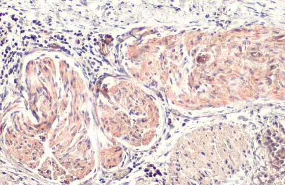
BCA1 antibody [N3C3] detects BCA1 protein by immunohistochemical analysis. Sample: Paraffin-embedded human esophageal carcinoma. BCA1 stained by BCA1 antibody [N3C3] (GTX108471) diluted at 1:500. Antigen Retrieval: Citrate buffer, pH 6.0, 15 min
![BCA1 antibody [N3C3] detects BCA1 protein at cytoplasm by immunohistochemical analysis. Sample: Paraffin-embedded human esophageal carcinoma. BCA1 stained by BCA1 antibody [N3C3] (GTX108471) diluted at 1:500. Antigen Retrieval: Citrate buffer, pH 6.0, 15 min](https://www.genetex.com/upload/website/prouct_img/normal/GTX108471/GTX108471_44762_20220819_IHC-P_22090701_911.webp "BCA1 antibody [N3C3] detects BCA1 protein at cytoplasm by immunohistochemical analysis. Sample: Paraffin-embedded human esophageal carcinoma. BCA1 stained by BCA1 antibody [N3C3] (GTX108471) diluted at 1:500. Antigen Retrieval: Citrate buffer, pH 6.0, 15 min")
![BCA1 antibody [N3C3] detects BCA1 protein at cell membrane and cytoplasm by immunohistochemical analysis. Sample: Paraffin-embedded human esophageal carcinoma. BCA1 stained by BCA1 antibody [N3C3] (GTX108471) diluted at 1:100. Antigen Retrieval: Citrate buffer, pH 6.0, 15 min](https://www.genetex.com/upload/website/prouct_img/normal/GTX108471/GTX108471_44174_20210205_IHC-P_w_23060120_923.webp "BCA1 antibody [N3C3] detects BCA1 protein at cell membrane and cytoplasm by immunohistochemical analysis. Sample: Paraffin-embedded human esophageal carcinoma. BCA1 stained by BCA1 antibody [N3C3] (GTX108471) diluted at 1:100. Antigen Retrieval: Citrate buffer, pH 6.0, 15 min")
![BCA1 antibody [N3C3] detects secreted BCA1 protein by immunofluorescent analysis. Sample: HepG2 cells were fixed in 4% paraformaldehyde at RT for 15 min. Green: BCA1 protein stained by BCA1 antibody [N3C3] (GTX108471) diluted at 1:500. Blue: Hoechst 33342 staining. Scale bar = 10 μm.](https://www.genetex.com/upload/website/prouct_img/normal/GTX108471/GTX108471_42844_20170621_IFA_w_23060120_204.webp "BCA1 antibody [N3C3] detects secreted BCA1 protein by immunofluorescent analysis. Sample: HepG2 cells were fixed in 4% paraformaldehyde at RT for 15 min. Green: BCA1 protein stained by BCA1 antibody [N3C3] (GTX108471) diluted at 1:500. Blue: Hoechst 33342 staining. Scale bar = 10 μm.")
![BCA1 antibody [N3C3] detects BCA1 protein at cytoplasm by immunohistochemical analysis. Sample: Paraffin-embedded human esophageal carcinoma. BCA1 stained by BCA1 antibody [N3C3] (GTX108471) diluted at 1:500. Antigen Retrieval: Citrate buffer, pH 6.0, 15 min](https://www.genetex.com/upload/website/prouct_img/normal/GTX108471/GTX108471_43852_20201127_IHC-P_w_23060120_929.webp "BCA1 antibody [N3C3] detects BCA1 protein at cytoplasm by immunohistochemical analysis. Sample: Paraffin-embedded human esophageal carcinoma. BCA1 stained by BCA1 antibody [N3C3] (GTX108471) diluted at 1:500. Antigen Retrieval: Citrate buffer, pH 6.0, 15 min")
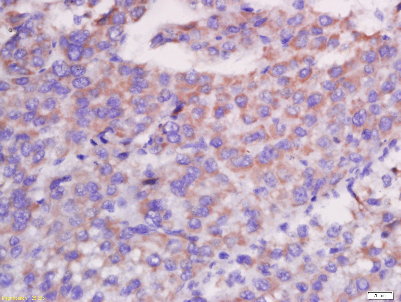
![BCA1 antibody [N3C3] detects BCA1 protein at cytoplasm in human esophageal carcinoma by immunohistochemical analysis. Sample: Paraffin-embedded human esophageal carcinoma. BCA1 antibody [N3C3] (GTX108471) diluted at 1:500.
Antigen Retrieval: Citrate buffer, pH 6.0, 15 min](https://www.genetex.com/upload/website/prouct_img/normal/GTX108471/GTX108471_42935_20170929_IHC-P_w_23060120_548.webp "BCA1 antibody [N3C3] detects BCA1 protein at cytoplasm in human esophageal carcinoma by immunohistochemical analysis. Sample: Paraffin-embedded human esophageal carcinoma. BCA1 antibody [N3C3] (GTX108471) diluted at 1:500.
Antigen Retrieval: Citrate buffer, pH 6.0, 15 min")
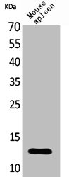
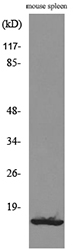
![Non-transfected (–) and transfected (+) 293T whole cell extracts (30 μg) were separated by 15% SDS-PAGE, and the membrane was blotted with BCA1 antibody [N3C3] (GTX108471) diluted at 1:10000. The HRP-conjugated anti-rabbit IgG antibody (GTX213110-01) was used to detect the primary antibody.](https://www.genetex.com/upload/website/prouct_img/normal/GTX108471/GTX108471_44762_20220805_WB_B_24122400_142.webp "Non-transfected (–) and transfected (+) 293T whole cell extracts (30 μg) were separated by 15% SDS-PAGE, and the membrane was blotted with BCA1 antibody [N3C3] (GTX108471) diluted at 1:10000. The HRP-conjugated anti-rabbit IgG antibody (GTX213110-01) was used to detect the primary antibody.")
BCA1 antibody [N3C3] detects BCA1 protein by immunohistochemical analysis. Sample: Paraffin-embedded human esophageal carcinoma. BCA1 stained by BCA1 antibody [N3C3] (GTX108471) diluted at 1:500. Antigen Retrieval: Citrate buffer, pH 6.0, 15 min
BCA1 antibody [N3C3]
GTX108471
ApplicationsImmunoFluorescence, Western Blot, ImmunoCytoChemistry, ImmunoHistoChemistry, ImmunoHistoChemistry Paraffin
Product group Antibodies
ReactivityHuman
TargetCXCL13
Overview
- SupplierGeneTex
- Product NameBCA1 antibody [N3C3]
- Delivery Days Customer9
- Application Supplier NoteWB: 1:1000-1:10000. ICC/IF: 1:100-1:1000. IHC-P: 1:100-1:1000. *Optimal dilutions/concentrations should be determined by the researcher.Not tested in other applications.
- ApplicationsImmunoFluorescence, Western Blot, ImmunoCytoChemistry, ImmunoHistoChemistry, ImmunoHistoChemistry Paraffin
- CertificationResearch Use Only
- ClonalityPolyclonal
- Concentration1.21 mg/ml
- ConjugateUnconjugated
- Gene ID10563
- Target nameCXCL13
- Target descriptionC-X-C motif chemokine ligand 13
- Target synonymsANGIE, ANGIE2, BCA-1, BCA1, BLC, BLR1L, SCYB13, C-X-C motif chemokine 13, B-cell chemoattractant, B-cell-attracting chemokine 1, B-cell-homing chemokine (ligand for Burkitt's lymphoma receptor-1), B-lymphocyte chemoattractant, CXC chemokine BLC, b cell-attracting chemokine 1, b lymphocyte chemoattractant, chemokine (C-X-C motif) ligand 13 (B-cell chemoattractant), small inducible cytokine B subfamily (Cys-X-Cys motif), member 13 (B-cell chemoattractant), small-inducible cytokine B13
- HostRabbit
- IsotypeIgG
- Protein IDO43927
- Protein NameC-X-C motif chemokine 13
- Scientific DescriptionB lymphocyte chemoattractant, independently cloned and named Angie, is a CXC chemokine strongly expressed in the follicles of the spleen, lymph nodes, and Peyers patches. It preferentially promotes the migration of B lymphocytes (compared to T cells and macrophages), apparently by stimulating calcium influx into, and chemotaxis of, cells expressing Burkitts lymphoma receptor 1 (BLR-1). It may therefore function in the homing of B lymphocytes to follicles. [provided by RefSeq]
- ReactivityHuman
- Storage Instruction-20°C or -80°C,2°C to 8°C
- UNSPSC41116161
Datasheet
Related products
Product group Antibodies
CXCL13 AntibodyCSB-PA005137
ApplicationsWestern Blot, ELISA
ReactivityHuman
TargetCXCL13
- SizePrice
Product group Antibodies
Anti-CXCL13 AntibodyA100583
ApplicationsWestern Blot, ELISA
ReactivityHuman
- SizePrice

Product group Antibodies
Anti-CXCL13 AntibodyAMAB91629
ApplicationsImmunoHistoChemistry
ReactivityHuman
TargetCXCL13
- SizePrice

Product group Antibodies
Anti-BCA1/CXCL13 Antibody Picoband(r)A02678-1-CARRIER-FREE
ApplicationsFlow Cytometry, Western Blot, ImmunoHistoChemistry
ReactivityHuman
TargetCXCL13
- SizePrice
Product group Antibodies

BLC / CXCL13 Antibody (aa22-109)LS-C293019
ApplicationsWestern Blot
ReactivityMouse
TargetCXCL13
- SizePrice
Product group Antibodies
References
CXCL13 Polyclonal AntibodyBS-2553R
ApplicationsImmunoFluorescence, Western Blot, ELISA, ImmunoCytoChemistry, ImmunoHistoChemistry, ImmunoHistoChemistry Frozen, ImmunoHistoChemistry Paraffin
ReactivityCanine, Human
TargetCXCL13
- SizePrice
Product group Antibodies
ApplicationsWestern Blot, ImmunoHistoChemistry
TargetCXCL13
- SizePrice
![BCA1 antibody [HL2172] detects BCA1 protein by immunohistochemical analysis. Sample: Paraffin-embedded human Clear Cell Renal Cell Carcinoma (ccRCC). BCA1 stained by BCA1 antibody [HL2172] (GTX638176) diluted at 1:100. Antigen Retrieval: Tris-EDTA buffer, pH 8.0, 15 min](https://www.genetex.com/upload/website/prouct_img/normal/GTX638176/GTX638176_T-44935_20230310_IHC-P_23041023_880.webp)
Product group Antibodies
BCA1 antibody [HL2172]GTX638176
ApplicationsWestern Blot, ImmunoHistoChemistry, ImmunoHistoChemistry Paraffin
ReactivityHuman
TargetCXCL13
- SizePrice
Product group Antibodies
BCA1 antibody [12A2]GTX52665
ApplicationsWestern Blot, Neutralisation/Blocking
ReactivityHuman
TargetCXCL13
- SizePrice