

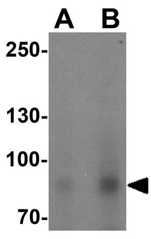
WB analysis of HeLa cell lysate using GTX17178 BCAR3 antibody. Working concentration : (A) 1 and (B) 2 μg/ml

WB analysis of HeLa cell lysate using GTX17178 BCAR3 antibody. Working concentration : (A) 1 and (B) 2 μg/ml
BCAR3 antibody
GTX17178
ApplicationsWestern Blot, ELISA, ImmunoHistoChemistry, ImmunoHistoChemistry Paraffin
Product group Antibodies
ReactivityHuman, Mouse, Rat
TargetBCAR3
Overview
- SupplierGeneTex
- Product NameBCAR3 antibody
- Delivery Days Customer9
- Application Supplier NoteWB: 1 - 2 microg/mL. IHC-P: 20 microg/mL. *Optimal dilutions/concentrations should be determined by the researcher.Not tested in other applications.
- ApplicationsWestern Blot, ELISA, ImmunoHistoChemistry, ImmunoHistoChemistry Paraffin
- CertificationResearch Use Only
- ClonalityPolyclonal
- Concentration1 mg/ml
- ConjugateUnconjugated
- Gene ID8412
- Target nameBCAR3
- Target descriptionBCAR3 adaptor protein, NSP family member
- Target synonymsAND-34, MIG7, NSP2, SH2D3B, breast cancer anti-estrogen resistance protein 3, BCAR3, NSP family adaptor protein, SH2 domain-containing protein 3B, breast cancer anti-estrogen resistance 3, breast cancer antiestrogen resistance 3 protein, dJ1033H22.2 (breast cancer anti-estrogen resistance 3), epididymis secretory sperm binding protein, mig-7, migration inducting gene-7, novel SH2-containing protein 2
- HostRabbit
- IsotypeIgG
- Protein IDO75815
- Protein NameBreast cancer anti-estrogen resistance protein 3
- Scientific DescriptionBreast tumors are initially dependent on estrogens for growth and progression and can be inhibited by anti-estrogens such as tamoxifen. However, breast cancers progress to become anti-estrogen resistant. Breast cancer anti-estrogen resistance gene 3 was identified in the search for genes involved in the development of estrogen resistance. The gene encodes a component of intracellular signal transduction that causes estrogen-independent proliferation in human breast cancer cells. The protein contains a putative src homology 2 (SH2) domain, a hall mark of cellular tyrosine kinase signaling molecules, and is partly homologous to the cell division cycle protein CDC48. Multiple transcript variants encoding different isoforms have been found for this gene. [provided by RefSeq, May 2012]
- ReactivityHuman, Mouse, Rat
- Storage Instruction-20°C or -80°C,2°C to 8°C
- UNSPSC41116161
Datasheet
Related products
Product group Antibodies
Anti-BCAR3 AntibodyA99678
ApplicationsWestern Blot, ELISA, ImmunoHistoChemistry
ReactivityHuman
- SizePrice
Product group Antibodies

Goat anti-BCAR3EB05071
ApplicationsWestern Blot, ELISA
ReactivityBovine, Canine, Human, Mouse, Porcine
TargetBCAR3
- SizePrice
Product group Antibodies
ApplicationsImmunoPrecipitation, Western Blot, ImmunoCytoChemistry, ImmunoHistoChemistry
ReactivityMouse, Rat
TargetBCAR3
- SizePrice
Product group Antibodies
BCAR3 AntibodyCSB-PA060303
ApplicationsImmunoFluorescence, Western Blot, ELISA, ImmunoHistoChemistry
ReactivityHuman
TargetBCAR3
- SizePrice
Product group Antibodies
BCAR3 antibody, C-termGTX89796
ApplicationsWestern Blot
ReactivityHuman, Mouse
TargetBCAR3
- SizePrice
Product group Antibodies
Anti-BCAR3 AntibodyHPA014858
ApplicationsWestern Blot, ImmunoCytoChemistry, ImmunoHistoChemistry
ReactivityHuman
TargetBCAR3
- SizePrice
Product group Antibodies

BCAR3 Antibody (aa25-75)LS-C288459
ApplicationsImmunoPrecipitation, Western Blot
ReactivityHuman
TargetBCAR3
- SizePrice

Product group Antibodies
Anti-BCAR3 Antibody Picoband(r)PB9846-CARRIER-FREE
ApplicationsWestern Blot, ImmunoHistoChemistry
ReactivityHuman, Mouse, Rat
TargetBCAR3
- SizePrice