

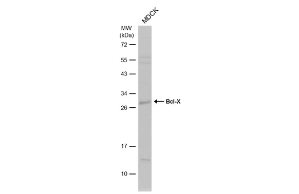
Whole cell extract (30 μg) was separated by 12% SDS-PAGE, and the membrane was blotted with Bcl-X antibody (GTX105661) diluted at 1:1000. The HRP-conjugated anti-rabbit IgG antibody (GTX213110-01) was used to detect the primary antibody, and the signal was developed with Trident ECL plus-Enhanced.
 antibody at 1:500 dilution.
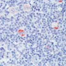
Antigen Retrieval: Trilogy? (EDTA based, pH 8.0) buffer, 15min")
 were separated by SDS-PAGE, and the membrane was blotted with Bcl-X antibody (GTX105661) diluted at 1:1000. The HRP-conjugated anti-rabbit IgG antibody (GTX213110-01) was used to detect the primary antibody.")
 antibody at 1:500 dilution.")
![Bcl-X antibody detects Bcl-X protein at cytoplasm by immunofluorescent analysis. Sample: SK-N-SH cells were fixed in 4% paraformaldehyde at RT for 15 min. Green: Bcl-X protein stained by Bcl-X antibody (GTX105661) diluted at 1:200. Red: alpha Tubulin, a cytoskeleton marker, stained by alpha Tubulin antibody [GT114] (GTX628802) diluted at 1:1000. Blue: Hoechst 33342 staining.](https://www.genetex.com/upload/website/prouct_img/normal/GTX105661/GTX105661_40051_20150410_IFA_w_23060120_931.webp "Bcl-X antibody detects Bcl-X protein at cytoplasm by immunofluorescent analysis. Sample: SK-N-SH cells were fixed in 4% paraformaldehyde at RT for 15 min. Green: Bcl-X protein stained by Bcl-X antibody (GTX105661) diluted at 1:200. Red: alpha Tubulin, a cytoskeleton marker, stained by alpha Tubulin antibody [GT114] (GTX628802) diluted at 1:1000. Blue: Hoechst 33342 staining.")
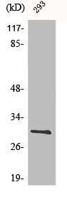
. Western blot analysis was performed using Bcl-X antibody (GTX105661) diluted at 1:500. EasyBlot anti-Rabbit IgG (GTX221666-01) was used as a secondary reagent.")
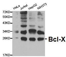
 were separated by 12% SDS-PAGE, and the membrane was blotted with Bcl-X antibody (GTX105661) diluted at 1:1000. The HRP-conjugated anti-rabbit IgG antibody (GTX213110-01) was used to detect the primary antibody.")
 A:NIH-3T3 12% SDS PAGE GTX105661 diluted at 1:1000 The HRP-conjugated anti-rabbit IgG antibody (GTX213110-01) was used to detect the primary antibody.")
 dilution: 1:1000 The HRP-conjugated anti-rabbit IgG antibody (GTX213110-01) was used to detect the primary antibody.")
 diluted at 1:500.
Antigen Retrieval: Trilogy? (EDTA based, pH 8.0) buffer, 15min")
Whole cell extract (30 μg) was separated by 12% SDS-PAGE, and the membrane was blotted with Bcl-X antibody (GTX105661) diluted at 1:1000. The HRP-conjugated anti-rabbit IgG antibody (GTX213110-01) was used to detect the primary antibody, and the signal was developed with Trident ECL plus-Enhanced.
Bcl-X antibody
GTX105661
ApplicationsImmunoFluorescence, ImmunoPrecipitation, Western Blot, ImmunoCytoChemistry, ImmunoHistoChemistry, ImmunoHistoChemistry Paraffin
Product group Antibodies
ReactivityCanine, Human, Mouse, Rat
TargetBCL2L1
Overview
- SupplierGeneTex
- Product NameBcl-X antibody
- Delivery Days Customer9
- Application Supplier NoteWB: 1:500-1:3000. ICC/IF: 1:100-1:1000. IHC-P: 1:100-1:1000. IP: 1:100-1:500. *Optimal dilutions/concentrations should be determined by the researcher.Not tested in other applications.
- ApplicationsImmunoFluorescence, ImmunoPrecipitation, Western Blot, ImmunoCytoChemistry, ImmunoHistoChemistry, ImmunoHistoChemistry Paraffin
- CertificationResearch Use Only
- ClonalityPolyclonal
- Concentration0.45 mg/ml
- ConjugateUnconjugated
- Gene ID598
- Target nameBCL2L1
- Target descriptionBCL2 like 1
- Target synonymsBCL-XL/S, BCL2L, BCLX, Bcl-X, PPP1R52, bcl-2-like protein 1, apoptosis regulator Bcl-X, protein phosphatase 1, regulatory subunit 52
- HostRabbit
- IsotypeIgG
- Protein IDQ07817
- Protein NameBcl-2-like protein 1
- Scientific DescriptionThe protein encoded by this gene belongs to the BCL-2 protein family. BCL-2 family members form hetero- or homodimers and act as anti- or pro-apoptotic regulators that are involved in a wide variety of cellular activities. The proteins encoded by this gene are located at the outer mitochondrial membrane, and have been shown to regulate outer mitochondrial membrane channel (VDAC) opening. VDAC regulates mitochondrial membrane potential, and thus controls the production of reactive oxygen species and release of cytochrome C by mitochondria, both of which are the potent inducers of cell apoptosis. Two alternatively spliced transcript variants, which encode distinct isoforms, have been reported. The longer isoform acts as an apoptotic inhibitor and the shorter form acts as an apoptotic activator. [provided by RefSeq]
- ReactivityCanine, Human, Mouse, Rat
- Storage Instruction-20°C or -80°C,2°C to 8°C
- UNSPSC41116161
Datasheet
Related products
Product group Antibodies
Anti-Bcl-X AntibodyA29719
ApplicationsWestern Blot, ImmunoHistoChemistry
ReactivityHuman, Mouse, Rat
- SizePrice
Product group Antibodies
Anti-BCL2L1 Antibody144-00209
ApplicationsImmunoFluorescence, ImmunoPrecipitation, Western Blot
ReactivityHuman, Mouse, Rat
TargetBCL2L1
- SizePrice

Product group Antibodies
Anti-Bcl-XL/BCL2L1 Antibody Picoband(r)A00181-CARRIER-FREE
ApplicationsFlow Cytometry, Western Blot
ReactivityHuman, Mouse, Rat
TargetBCL2L1
- SizePrice

Product group Antibodies
References
BCL2L1 Polyclonal AntibodyBS-1336R
ApplicationsFlow Cytometry, ImmunoFluorescence, Western Blot, ELISA, ImmunoCytoChemistry, ImmunoHistoChemistry, ImmunoHistoChemistry Frozen, ImmunoHistoChemistry Paraffin
ReactivityCanine, Equine, Human, Mouse, Porcine, Rat, Sheep
TargetBCL2L1
- SizePrice
Product group Antibodies
BCL2L1 AntibodyCSB-PA000986
ApplicationsImmunoFluorescence, Western Blot, ELISA, ImmunoHistoChemistry
ReactivityHuman, Mouse, Rat
TargetBCL2L1
- SizePrice
Product group Antibodies
Bcl2L1 Polyclonal AntibodyCAC11764
ApplicationsImmunoFluorescence, Western Blot, ELISA, ImmunoHistoChemistry
ReactivityMouse
TargetBCL2L1
- SizePrice
Product group Antibodies
Bcl-X antibody [2H12]GTX23193
ApplicationsFlow Cytometry, ImmunoFluorescence, Western Blot, ImmunoCytoChemistry, ImmunoHistoChemistry, ImmunoHistoChemistry Paraffin
ReactivityHuman, Mouse, Porcine, Rat
TargetBCL2L1
- SizePrice
Product group Antibodies
Bcl-X antibody [2H12]GTX25713
ApplicationsWestern Blot, ImmunoHistoChemistry
ReactivityHuman, Mouse
TargetBCL2L1
- SizePrice
Product group Antibodies
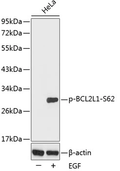
Bcl-X (phospho Ser62) antibodyGTX30085
ApplicationsWestern Blot
ReactivityHuman
TargetBCL2L1
- SizePrice
![WB analysis of various samples using GTX01535 Bcl-XL antibody [GT1227]. Dilution : 1:1000 Loading : 25 μg](https://www.genetex.com/upload/website/prouct_img/normal/GTX01535/GTX01535_20200508_WB_w_23053121_834.webp)
Product group Antibodies
Bcl-XL antibody [GT1227]GTX01535
ApplicationsWestern Blot
ReactivityHuman, Mouse, Rat
TargetBCL2L1
- SizePrice