

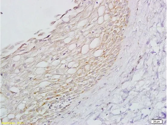
IHC-P analysis of skin of rat foot tissue using GTX04321 beta 1 Adrenergic Receptor antibody. Dilution : 1:200


IHC-P analysis of skin of rat foot tissue using GTX04321 beta 1 Adrenergic Receptor antibody. Dilution : 1:200
beta 1 Adrenergic Receptor antibody
GTX04321
ApplicationsFlow Cytometry, Western Blot, ImmunoHistoChemistry, ImmunoHistoChemistry Paraffin
Product group Antibodies
ReactivityHuman, Mouse, Rat
TargetADRB1
Overview
- SupplierGeneTex
- Product Namebeta 1 Adrenergic Receptor antibody
- Delivery Days Customer9
- Application Supplier NoteWB: 1:300-1:5000. IHC-P: 1:200-1:400. FCM: 1:20-1:100. *Optimal dilutions/concentrations should be determined by the researcher.Not tested in other applications.
- ApplicationsFlow Cytometry, Western Blot, ImmunoHistoChemistry, ImmunoHistoChemistry Paraffin
- CertificationResearch Use Only
- ClonalityPolyclonal
- Concentration1 mg/ml
- ConjugateUnconjugated
- Gene ID153
- Target nameADRB1
- Target descriptionadrenoceptor beta 1
- Target synonymsADRB1R, B1AR, BETA1AR, FNSS2, RHR, beta-1 adrenergic receptor, adrenergic, beta-1-, receptor, beta-1 adrenoceptor, beta-1 adrenoreceptor
- HostRabbit
- IsotypeIgG
- Protein IDP08588
- Protein NameBeta-1 adrenergic receptor
- Scientific DescriptionThe adrenergic receptors (subtypes alpha 1, alpha 2, beta 1, and beta 2) are a prototypic family of guanine nucleotide binding regulatory protein-coupled receptors that mediate the physiological effects of the hormone epinephrine and the neurotransmitter norepinephrine. Specific polymorphisms in this gene have been shown to affect the resting heart rate and can be involved in heart failure. [provided by RefSeq, Jul 2008]
- ReactivityHuman, Mouse, Rat
- Storage Instruction-20°C or -80°C,2°C to 8°C
- UNSPSC41116161
Datasheet
Related products

Product group Antibodies
ADRB1 AntibodyCSB-PA000937
ApplicationsImmunoFluorescence, Western Blot, ELISA, ImmunoHistoChemistry
ReactivityHuman, Mouse, Rat
TargetADRB1
- SizePrice

Product group Antibodies
Anti-ADRB1 Antibody Picoband(r)A00675-2-CARRIER-FREE
ApplicationsWestern Blot
ReactivityMouse, Rat
TargetADRB1
- SizePrice
Product group Antibodies
ApplicationsImmunoFluorescence, ELISA, ImmunoHistoChemistry
ReactivityHuman
- SizePrice
Product group Antibodies
References

Goat anti-ADRB1EB07133
ApplicationsImmunoFluorescence, ELISA, ImmunoHistoChemistry
ReactivityCanine, Human, Mouse, Rat
TargetADRB1
- SizePrice
Product group Antibodies

ADRB1 Antibody (Internal)LS-C368864
ApplicationsImmunoFluorescence, Western Blot, ImmunoCytoChemistry, ImmunoHistoChemistry, ImmunoHistoChemistry Paraffin
ReactivityHuman
TargetADRB1
- SizePrice
Product group Antibodies
ADRB1 Polyclonal AntibodyCAC14630
ApplicationsImmunoFluorescence, Western Blot, ELISA, ImmunoHistoChemistry
ReactivityMouse
TargetADRB1
- SizePrice
![Non-transfected (–) and transfected (+) unboiled MDA-MB-231 whole cell extracts (30 μg) were separated by 10% SDS-PAGE, and the membrane was blotted with beta 1 Adrenergic Receptor antibody [HL3678] (GTX641706) diluted at 1:1000. The HRP-conjugated anti-rabbit IgG antibody (GTX213110-01) was used to detect the primary antibody.](https://www.genetex.com/upload/website/prouct_img/normal/GTX641706/GTX641706_T-45649_20250207_WB_shRNA_watermark_25021320_678.webp)
Product group Antibodies
ApplicationsFlow Cytometry, Western Blot
ReactivityHuman
TargetADRB1
- SizePrice
Product group Antibodies
ApplicationsImmunoFluorescence, ImmunoCytoChemistry, ImmunoHistoChemistry, ImmunoHistoChemistry Paraffin
ReactivityHuman
TargetADRB1
- SizePrice
Product group Antibodies
References
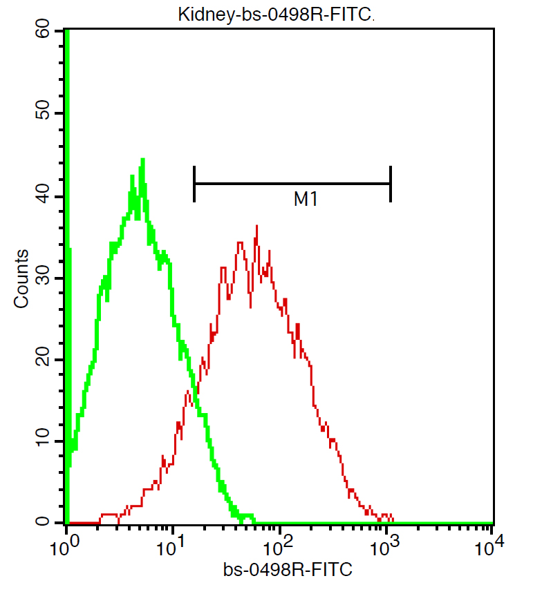
ADRB1 Polyclonal AntibodyBS-0498R
ApplicationsFlow Cytometry, ImmunoFluorescence, Western Blot, ELISA, ImmunoCytoChemistry, ImmunoHistoChemistry, ImmunoHistoChemistry Frozen, ImmunoHistoChemistry Paraffin
ReactivityCanine, Human, Mouse, Porcine, Rat
TargetADRB1
- SizePrice
Product group Antibodies
Anti-ADRB1 Antibody144-64286
ApplicationsWestern Blot
ReactivityHuman, Mouse, Rat
TargetADRB1
- SizePrice