


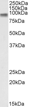
Wild-type (WT) and CTNNB1 knockout (KO) 293T cell extracts (30 μg) were separated by 7.5% SDS-PAGE, and the membrane was blotted with beta Catenin antibody [HL1753] (GTX637403) diluted at 1:1000. The HRP-conjugated anti-rabbit IgG antibody (GTX213110-01) was used to detect the primary antibody, and the signal was developed with Trident ECL plus-Enhanced.
![Various whole cell extracts (30 μg) were separated by 7.5% SDS-PAGE, and the membrane was blotted with beta Catenin antibody [HL1753] (GTX637403) diluted at 1:1000. The HRP-conjugated anti-rabbit IgG antibody (GTX213110-01) was used to detect the primary antibody.](https://www.genetex.com/upload/website/prouct_img/normal/GTX637403/GTX637403_44858_20221111_WB_22111518_821.webp "Various whole cell extracts (30 μg) were separated by 7.5% SDS-PAGE, and the membrane was blotted with beta Catenin antibody [HL1753] (GTX637403) diluted at 1:1000. The HRP-conjugated anti-rabbit IgG antibody (GTX213110-01) was used to detect the primary antibody.")
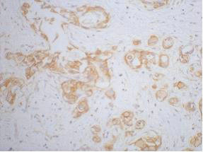
![beta Catenin antibody [HL1753] detects beta Catenin protein at cell membrane by immunohistochemical analysis. Sample: Paraffin-embedded mouse tissue. beta Catenin stained by beta Catenin antibody [HL1753] (GTX637403) diluted at 1:100. Antigen Retrieval: Citrate buffer, pH 6.0, 15 min](https://www.genetex.com/upload/website/prouct_img/normal/GTX637403/GTX637403_T-44788_20221202_IHC-P_M_22121123_278.webp "beta Catenin antibody [HL1753] detects beta Catenin protein at cell membrane by immunohistochemical analysis. Sample: Paraffin-embedded mouse tissue. beta Catenin stained by beta Catenin antibody [HL1753] (GTX637403) diluted at 1:100. Antigen Retrieval: Citrate buffer, pH 6.0, 15 min")
![beta Catenin antibody [HL1753] detects beta Catenin protein at cell membrane by immunohistochemical analysis. Sample: Paraffin-embedded cat mammary gland. beta Catenin stained by beta Catenin antibody [HL1753] (GTX637403) diluted at 1:100. Antigen Retrieval: Citrate buffer, pH 6.0, 15 min](https://www.genetex.com/upload/website/prouct_img/normal/GTX637403/GTX637403_44858_20230203_IHC-P_Cat_23020621_168.webp "beta Catenin antibody [HL1753] detects beta Catenin protein at cell membrane by immunohistochemical analysis. Sample: Paraffin-embedded cat mammary gland. beta Catenin stained by beta Catenin antibody [HL1753] (GTX637403) diluted at 1:100. Antigen Retrieval: Citrate buffer, pH 6.0, 15 min")
![beta Catenin antibody [HL1753] detects beta Catenin protein at cell membrane by immunohistochemical analysis. Sample: Paraffin-embedded dog mammary gland. beta Catenin stained by beta Catenin antibody [HL1753] (GTX637403) diluted at 1:100. Antigen Retrieval: Citrate buffer, pH 6.0, 15 min](https://www.genetex.com/upload/website/prouct_img/normal/GTX637403/GTX637403_44858_20230203_IHC-P_Dog_23020621_922.webp "beta Catenin antibody [HL1753] detects beta Catenin protein at cell membrane by immunohistochemical analysis. Sample: Paraffin-embedded dog mammary gland. beta Catenin stained by beta Catenin antibody [HL1753] (GTX637403) diluted at 1:100. Antigen Retrieval: Citrate buffer, pH 6.0, 15 min")
![beta Catenin antibody [HL1753] detects beta Catenin protein at cell membrane and cell junction by immunofluorescent analysis. Sample: A431 cells were fixed in 4% paraformaldehyde at RT for 15 min. Green: beta Catenin stained by beta Catenin antibody [HL1753] (GTX637403) diluted at 1:500. Blue: Fluoroshield with DAPI (GTX30920).](https://www.genetex.com/upload/website/prouct_img/normal/GTX637403/GTX637403_44858_20230119_ICC_IF_23021401_687.webp "beta Catenin antibody [HL1753] detects beta Catenin protein at cell membrane and cell junction by immunofluorescent analysis. Sample: A431 cells were fixed in 4% paraformaldehyde at RT for 15 min. Green: beta Catenin stained by beta Catenin antibody [HL1753] (GTX637403) diluted at 1:500. Blue: Fluoroshield with DAPI (GTX30920).")
Wild-type (WT) and CTNNB1 knockout (KO) 293T cell extracts (30 μg) were separated by 7.5% SDS-PAGE, and the membrane was blotted with beta Catenin antibody [HL1753] (GTX637403) diluted at 1:1000. The HRP-conjugated anti-rabbit IgG antibody (GTX213110-01) was used to detect the primary antibody, and the signal was developed with Trident ECL plus-Enhanced.
beta Catenin antibody [HL1753]
GTX637403
ApplicationsImmunoFluorescence, Western Blot, ImmunoCytoChemistry, ImmunoHistoChemistry, ImmunoHistoChemistry Paraffin
Product group Antibodies
ReactivityCanine, Feline, Human, Mouse
TargetCTNNB1
Overview
- SupplierGeneTex
- Product Namebeta Catenin antibody [HL1753]
- Delivery Days Customer9
- Application Supplier NoteWB: 1:500-1:3000. *Optimal dilutions/concentrations should be determined by the researcher.Not tested in other applications.
- ApplicationsImmunoFluorescence, Western Blot, ImmunoCytoChemistry, ImmunoHistoChemistry, ImmunoHistoChemistry Paraffin
- CertificationResearch Use Only
- ClonalityMonoclonal
- Clone IDHL1753
- Concentration1 mg/ml
- ConjugateUnconjugated
- Gene ID1499
- Target nameCTNNB1
- Target descriptioncatenin beta 1
- Target synonymsCTNNB, EVR7, MRD19, NEDSDV, armadillo, catenin beta-1, catenin (cadherin-associated protein), beta 1, 88kDa
- HostRabbit
- IsotypeIgG
- Protein IDP35222
- Protein NameCatenin beta-1
- Scientific DescriptionThe protein encoded by this gene is part of a complex of proteins that constitute adherens junctions (AJs). AJs are necessary for the creation and maintenance of epithelial cell layers by regulating cell growth and adhesion between cells. The encoded protein also anchors the actin cytoskeleton and may be responsible for transmitting the contact inhibition signal that causes cells to stop dividing once the epithelial sheet is complete. Finally, this protein binds to the product of the APC gene, which is mutated in adenomatous polyposis of the colon. Mutations in this gene are a cause of colorectal cancer (CRC), pilomatrixoma (PTR), medulloblastoma (MDB), and ovarian cancer. Alternative splicing results in multiple transcript variants. [provided by RefSeq, Aug 2016]
- ReactivityCanine, Feline, Human, Mouse
- Storage Instruction-20°C or -80°C,2°C to 8°C
- UNSPSC41116161
Datasheet
Related products
Product group Antibodies
ReactivityHuman
TargetCTNNB1
- SizePrice
Product group Antibodies
Anti-beta Catenin AntibodyA121145
ApplicationsFlow Cytometry, ImmunoFluorescence, Western Blot, ELISA
ReactivityHuman
- SizePrice

Product group Antibodies
Anti-beta Catenin/CTNNB1 Antibody Picoband(r)A00004-CARRIER-FREE
ApplicationsFlow Cytometry, ImmunoFluorescence, Western Blot, ELISA, ImmunoCytoChemistry, ImmunoHistoChemistry
ReactivityHuman, Mouse, Rat
TargetCTNNB1
- SizePrice

Product group Antibodies
Anti-CTNNB1 AntibodyAMAB91209
ApplicationsWestern Blot, ImmunoCytoChemistry, ImmunoHistoChemistry
ReactivityHuman
TargetCTNNB1
- SizePrice
Product group Antibodies

Anti-beta catenin [15B8]Ab01655-1.1
ApplicationsFlow Cytometry, ImmunoFluorescence, Western Blot, ImmunoHistoChemistry, Other Application
ReactivityBovine, Canine, Chicken, Human, Mouse, Rat
TargetCTNNB1
- SizePrice
Product group Antibodies

ApplicationsWestern Blot, ImmunoHistoChemistry, ImmunoHistoChemistry Paraffin
ReactivityHuman, Mouse, Rat
TargetCTNNB1
- SizePrice
Product group Antibodies

ApplicationsFlow Cytometry, ImmunoFluorescence, Western Blot, ELISA
ReactivityBovine, Canine, Human, Mouse, Porcine, Rat
TargetCTNNB1
- SizePrice
Product group Antibodies
CTNNB1 Monoclonal AntibodyCSB-MA000223
ApplicationsWestern Blot, ELISA, ImmunoHistoChemistry
ReactivityHuman, Mouse, Rat
TargetCTNNB1
- SizePrice
Product group Antibodies
References
beta Catenin antibodyGTX26302
ApplicationsDot Blot, ImmunoFluorescence, ImmunoPrecipitation, Western Blot, ImmunoCytoChemistry, ImmunoHistoChemistry, ImmunoHistoChemistry Frozen, ImmunoHistoChemistry Paraffin
ReactivityFish, Human, Mouse, Porcine, Rat, Xenopus, Zebra Fish
TargetCTNNB1
- SizePrice
Product group Antibodies
Ctnnb1 Polyclonal AntibodyCAC07113
ApplicationsImmunoFluorescence, ImmunoPrecipitation, Western Blot, ELISA, ImmunoHistoChemistry
TargetCTNNB1
- SizePrice