

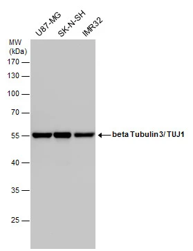
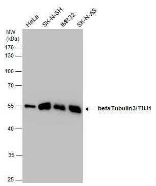
beta Tubulin 3/ TUJ1 antibody detects beta Tubulin 3/ TUJ1 protein by Western blot analysis. Various whole cell extracts (30 μg) were separated by 10% SDS-PAGE, and the membrane was blotted with beta Tubulin 3/ TUJ1 antibody (GTX631831) diluted by 1:1000.
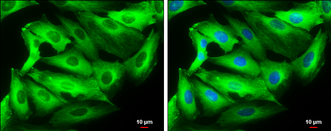
![beta Tubulin 3/ Tuj1 antibody [GT1338] detects beta Tubulin 3/ Tuj1 protein at cytoplasm by immunofluorescent analysis. Sample: DIV9 rat E18 primary cortical neurons were fixed in 4% paraformaldehyde at RT for 15 min. Green: beta Tubulin 3/ Tuj1 protein stained by beta Tubulin 3/ Tuj1 antibody [GT1338] (GTX631831) diluted at 1:500. Red: NeuN, stained by NeuN antibody (GTX132974) diluted at 1:1000. Blue: Fluoroshield with DAPI (GTX30920).](https://www.genetex.com/upload/website/prouct_img/normal/GTX631831/GTX631831_41883_20170503_IFA_R_w_23061202_541.webp "beta Tubulin 3/ Tuj1 antibody [GT1338] detects beta Tubulin 3/ Tuj1 protein at cytoplasm by immunofluorescent analysis. Sample: DIV9 rat E18 primary cortical neurons were fixed in 4% paraformaldehyde at RT for 15 min. Green: beta Tubulin 3/ Tuj1 protein stained by beta Tubulin 3/ Tuj1 antibody [GT1338] (GTX631831) diluted at 1:500. Red: NeuN, stained by NeuN antibody (GTX132974) diluted at 1:1000. Blue: Fluoroshield with DAPI (GTX30920).")
![Wild-type (WT) and beta Tubulin 3/ Tuj1 knockout (KO) HeLa cell extracts (30 μg) were separated by 10% SDS-PAGE, and the membrane was blotted with beta Tubulin 3/ Tuj1 antibody [GT1338] (GTX631831) diluted at 1:5000. The HRP-conjugated anti-mouse IgG antibody (GTX213111-01) was used to detect the primary antibody.](https://www.genetex.com/upload/website/prouct_img/normal/GTX631831/GTX631831_41883_20170309_WB_KO_watermark_w_23061202_280.webp "Wild-type (WT) and beta Tubulin 3/ Tuj1 knockout (KO) HeLa cell extracts (30 μg) were separated by 10% SDS-PAGE, and the membrane was blotted with beta Tubulin 3/ Tuj1 antibody [GT1338] (GTX631831) diluted at 1:5000. The HRP-conjugated anti-mouse IgG antibody (GTX213111-01) was used to detect the primary antibody.")
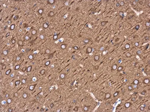
![beta Tubulin 3/ Tuj1 antibody [GT1338] detects beta Tubulin 3/ Tuj1 protein at cytoplasm by immunohistochemical analysis. Sample: Paraffin-embedded mouse E10.5 embryo. Red: beta Tubulin 3/ Tuj1 stained by beta Tubulin 3/ Tuj1 antibody [GT1338] (GTX631831) diluted at 1:500. Blue: Fluoroshield with DAPI (GTX30920).](https://www.genetex.com/upload/website/prouct_img/normal/GTX631831/GTX631831_41883_20180713_IHC-P-FL_M_w_23061202_874.webp "beta Tubulin 3/ Tuj1 antibody [GT1338] detects beta Tubulin 3/ Tuj1 protein at cytoplasm by immunohistochemical analysis. Sample: Paraffin-embedded mouse E10.5 embryo. Red: beta Tubulin 3/ Tuj1 stained by beta Tubulin 3/ Tuj1 antibody [GT1338] (GTX631831) diluted at 1:500. Blue: Fluoroshield with DAPI (GTX30920).")
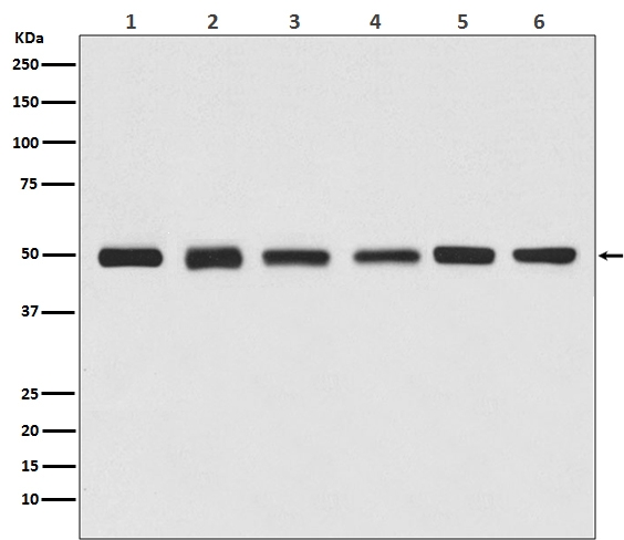
![Various tissue extracts (50 μg) were separated by 10% SDS-PAGE, and the membrane was blotted with beta Tubulin 3/ Tuj1 antibody [GT1338] (GTX631831) diluted at 1:10000. The HRP-conjugated anti-mouse IgG antibody (GTX213111-01) was used to detect the primary antibody.](https://www.genetex.com/upload/website/prouct_img/normal/GTX631831/GTX631831_41883_20170713_WB_M_R_w_23061202_292.webp "Various tissue extracts (50 μg) were separated by 10% SDS-PAGE, and the membrane was blotted with beta Tubulin 3/ Tuj1 antibody [GT1338] (GTX631831) diluted at 1:10000. The HRP-conjugated anti-mouse IgG antibody (GTX213111-01) was used to detect the primary antibody.")
beta Tubulin 3/ TUJ1 antibody detects beta Tubulin 3/ TUJ1 protein by Western blot analysis. Various whole cell extracts (30 μg) were separated by 10% SDS-PAGE, and the membrane was blotted with beta Tubulin 3/ TUJ1 antibody (GTX631831) diluted by 1:1000.
beta Tubulin 3/ Tuj1 antibody [GT1338]
GTX631831
ApplicationsImmunoFluorescence, Western Blot, ImmunoCytoChemistry, ImmunoHistoChemistry, ImmunoHistoChemistry Paraffin
Product group Antibodies
ReactivityHuman, Mouse, Rat
TargetTUBB3
Overview
- SupplierGeneTex
- Product Namebeta Tubulin 3/ Tuj1 antibody [GT1338]
- Delivery Days Customer9
- Application Supplier NoteWB: 1:500-1:20000. ICC/IF: 1:100-1:1000. IHC-P: 1:100-1:1000. *Optimal dilutions/concentrations should be determined by the researcher.Not tested in other applications.
- ApplicationsImmunoFluorescence, Western Blot, ImmunoCytoChemistry, ImmunoHistoChemistry, ImmunoHistoChemistry Paraffin
- CertificationResearch Use Only
- ClonalityMonoclonal
- Clone IDGT1338
- Concentration1.79 mg/ml
- ConjugateUnconjugated
- Gene ID10381
- Target nameTUBB3
- Target descriptiontubulin beta 3 class III
- Target synonymsCDCBM, CDCBM1, CFEOM3, CFEOM3A, FEOM3, TUBB4, beta-4, tubulin beta-3 chain, class III beta-tubulin, tubulin beta-4 chain, tubulin beta-III, tubulin, beta 3
- HostMouse
- IsotypeIgG2a
- Protein IDQ13509
- Protein NameTubulin beta-3 chain
- Scientific DescriptionThis gene encodes a class III member of the beta tubulin protein family. Beta tubulins are one of two core protein families (alpha and beta tubulins) that heterodimerize and assemble to form microtubules. This protein is primarily expressed in neurons and may be involved in neurogenesis and axon guidance and maintenance. Mutations in this gene are the cause of congenital fibrosis of the extraocular muscles type 3. Alternate splicing results in multiple transcript variants. A pseudogene of this gene is found on chromosome 6. [provided by RefSeq]
- ReactivityHuman, Mouse, Rat
- Storage Instruction-20°C or -80°C,2°C to 8°C
- UNSPSC12352203
Datasheet
Related products
Product group Antibodies

Anti-Beta-Tubulin [S11B]Ab00404-1.1
ApplicationsImmunoFluorescence, Western Blot, ELISA
ReactivityHuman
TargetTUBB3
- SizePrice
Product group Antibodies
Anti-TUBB3 Antibody144-64781
ApplicationsImmunoFluorescence, ImmunoPrecipitation, Western Blot
ReactivityHuman, Mouse, Rat
TargetTUBB3
- SizePrice
Product group Antibodies
References
ApplicationsWestern Blot
ReactivityHuman, Mouse, Rat
TargetTUBB3
- SizePrice
Product group Antibodies
References
beta Tubulin 3/ Tuj1 antibodyGTX129913
ApplicationsImmunoFluorescence, Western Blot, ImmunoCytoChemistry, ImmunoHistoChemistry, ImmunoHistoChemistry Frozen
ReactivityHuman, Mouse, Rat
TargetTUBB3
- SizePrice
Product group Antibodies
References
beta Tubulin 3/ Tuj1 antibodyGTX130245
ApplicationsFlow Cytometry, ImmunoFluorescence, ImmunoPrecipitation, Western Blot, ImmunoCytoChemistry, ImmunoHistoChemistry, ImmunoHistoChemistry Frozen, ImmunoHistoChemistry Paraffin
ReactivityHuman, Mouse, Rat
TargetTUBB3
- SizePrice
![ICC/IF analysis of SH-SY-5Y cells using GTX11314 beta Tubulin 3/ Tuj1 antibody [SDL.3D10] at 1:400(pink) with DAPI(blue). Cells were fixed and permeabilized with methanol followed by methanol:acetone.](https://www.genetex.com/upload/website/prouct_img/normal/GTX11314/GTX11314_20170605_ICCIF_w_23060500_832.webp)
Product group Antibodies
ApplicationsImmunoFluorescence, Western Blot, ELISA, ImmunoCytoChemistry
ReactivityBovine, Human, Porcine, Rat
TargetTUBB3
- SizePrice
Product group Antibodies
ApplicationsImmunoFluorescence, ImmunoPrecipitation, Western Blot, ImmunoCytoChemistry, ImmunoHistoChemistry, ImmunoHistoChemistry Frozen, ImmunoHistoChemistry Paraffin
ReactivityHuman, Mouse, Rat
TargetTUBB3
- SizePrice
![beta Tubulin 3/ TUJ1 antibody [GT11710] detects beta Tubulin 3/ TUJ1 protein by immunohistochemical analysis. Sample: Frozen sectioned E13.5 rat brain. Red: beta Tubulin 3/ TUJ1 protein stained by beta Tubulin 3/ TUJ1 antibody [GT11710] (GTX131836) diluted at 1:250. Blue: Fluoroshield with DAPI (GTX30920).](https://www.genetex.com/upload/website/prouct_img/normal/GTX631836/GTX631836_41855_20160808_2_IHC-Fr_w_23061202_557.webp)
Product group Antibodies
References
ApplicationsImmunoFluorescence, ImmunoPrecipitation, Western Blot, ImmunoCytoChemistry, ImmunoHistoChemistry, ImmunoHistoChemistry Frozen, ImmunoHistoChemistry Paraffin
ReactivityFish, Human, Mouse, Rat
TargetTUBB3
- SizePrice
![beta Tubulin 3/ Tuj1 antibody [HL1708] detects beta Tubulin 3/ Tuj1 protein at cytoskeleton by immunofluorescent analysis. Sample: DIV9 rat E18 primary hippocampal neuron cells were fixed in 4% paraformaldehyde at RT for 15 min. Green: beta Tubulin 3/ Tuj1 stained by beta Tubulin 3/ Tuj1 antibody [HL1708] (GTX637307) diluted at 1:250. Blue: Fluoroshield with DAPI (GTX30920).](https://www.genetex.com/upload/website/prouct_img/normal/GTX637307/GTX637307_T-44774_20221209_ICC_IF_R_22122018_744.webp)
Product group Antibodies
ApplicationsImmunoFluorescence, Western Blot, ImmunoCytoChemistry
ReactivityHuman, Mouse, Rat
TargetTUBB3
- SizePrice