

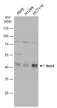
Various whole cell extracts (30 μg) were separated by 10% SDS-PAGE, and the membrane was blotted with Bmi1 antibody (GTX102020) diluted at 1:1000. The HRP-conjugated anti-rabbit IgG antibody (GTX213110-01) was used to detect the primary antibody.
 were separated by 10% SDS-PAGE, and the membrane was blotted with Bmi1 antibody (GTX102020) diluted at 1:1000. The HRP-conjugated anti-rabbit IgG antibody (GTX213110-01) was used to detect the primary antibody.")
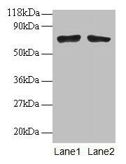
![Bmi1 antibody immunoprecipitates Bmi1 protein in IP experiments. IP samples: K562 whole cell extract A. 40 μg K562 whole cell extract B. Control with 4 μg of preimmune Rabbit IgG C. Immunoprecipitation of Bmi1 protein by 4 μg Bmi1 antibody (GTX102020) 5 % SDS-PAGE The immunoprecipitated Bmi1 protein was detected by Bmi1 antibody (GTX102020) diluted at 1:500. [EasyBlot anti-rabbit IgG (GTX221666-01) was used as a secondary reagent]](https://www.genetex.com/upload/website/prouct_img/normal/GTX102020/GTX102020_41101_IP_w_23060100_389.webp "Bmi1 antibody immunoprecipitates Bmi1 protein in IP experiments. IP samples: K562 whole cell extract A. 40 μg K562 whole cell extract B. Control with 4 μg of preimmune Rabbit IgG C. Immunoprecipitation of Bmi1 protein by 4 μg Bmi1 antibody (GTX102020) 5 % SDS-PAGE The immunoprecipitated Bmi1 protein was detected by Bmi1 antibody (GTX102020) diluted at 1:500. [EasyBlot anti-rabbit IgG (GTX221666-01) was used as a secondary reagent]")
Various whole cell extracts (30 μg) were separated by 10% SDS-PAGE, and the membrane was blotted with Bmi1 antibody (GTX102020) diluted at 1:1000. The HRP-conjugated anti-rabbit IgG antibody (GTX213110-01) was used to detect the primary antibody.
Bmi1 antibody
GTX102020
ApplicationsImmunoPrecipitation, Western Blot
Product group Antibodies
ReactivityHuman
TargetBMI1
Overview
- SupplierGeneTex
- Product NameBmi1 antibody
- Delivery Days Customer9
- Application Supplier NoteWB: 1:500-1:3000. IP: 1:100-1:500. *Optimal dilutions/concentrations should be determined by the researcher.Not tested in other applications.
- ApplicationsImmunoPrecipitation, Western Blot
- CertificationResearch Use Only
- ClonalityPolyclonal
- Concentration1 mg/ml
- ConjugateUnconjugated
- Gene ID648
- Target nameBMI1
- Target descriptionBMI1 proto-oncogene, polycomb ring finger
- Target synonymsFLVI2/BMI1, PCGF4, RNF51, flvi-2/bmi-1, polycomb complex protein BMI-1, B lymphoma Mo-MLV insertion region 1 homolog, BMI1 polycomb ring finger oncogene, BMI1 polycomb ring finger proto-oncogene, murine leukemia viral (bmi-1) oncogene homolog, polycomb group RING finger protein 4, polycomb group protein Bmi1, ring finger protein 51
- HostRabbit
- IsotypeIgG
- Protein IDP35226
- Protein NamePolycomb complex protein BMI-1
- Scientific DescriptionComponent of the Polycomb group (PcG) multiprotein PRC1 complex, a complex required to maintain the transcriptionally repressive state of many genes, including Hox genes, throughout development. PcG PRC1 complex acts via chromatin remodeling and modification of histones; it mediates monoubiquitination of histone H2A Lys-119, rendering chromatin heritably changed in its expressibility. In the PRC1 complex, it is required to stimulate the E3 ubiquitin-protein ligase activity of RNF2/RING2.
- ReactivityHuman
- Storage Instruction-20°C or -80°C,2°C to 8°C
- UNSPSC41116161
Datasheet
Related products

Product group Antibodies
Anti-Bmi1 AntibodyA85203
ApplicationsWestern Blot, ELISA, ImmunoHistoChemistry
ReactivityHuman
- SizePrice
Product group Antibodies
Anti-BMI1 Antibody144-00211
ApplicationsImmunoPrecipitation, Western Blot
ReactivityHuman
TargetBMI1
- SizePrice
Product group Antibodies

BMI1 / PCGF4 AntibodyLS-C831530
ApplicationsWestern Blot
ReactivityMouse
TargetBMI1
- SizePrice
Product group Antibodies

Goat anti-BMI1 (aa237-251)EB12495
ApplicationsWestern Blot, ELISA, ImmunoHistoChemistry
ReactivityCanine, Human, Mouse, Porcine
TargetBMI1
- SizePrice
Product group Antibodies
BMI1 Polyclonal AntibodyCAC14038
ApplicationsWestern Blot, ELISA, ImmunoHistoChemistry
TargetBMI1
- SizePrice
Product group Antibodies
BMI1 AntibodyCSB-PA03345A0RB
ApplicationsWestern Blot, ELISA, ImmunoHistoChemistry
ReactivityHuman
TargetBMI1
- SizePrice
Product group Antibodies
PCGF4/BMI1 Polyclonal AntibodyBS-2999R
ApplicationsFlow Cytometry, ImmunoFluorescence, Western Blot, ELISA, ImmunoCytoChemistry, ImmunoHistoChemistry, ImmunoHistoChemistry Frozen, ImmunoHistoChemistry Paraffin
ReactivityBovine, Canine, Chicken, Human, Mouse, Rabbit, Rat
TargetBMI1
- SizePrice
Product group Antibodies

Mouse anti Human BMI1MUB2004P
ApplicationsImmunoPrecipitation, Western Blot, ImmunoHistoChemistry, ImmunoHistoChemistry Frozen
ReactivityHuman, Mouse, Rabbit, Rat
TargetBMI1
- SizePrice
Product group Antibodies
Bmi1 antibodyGTX31296
ApplicationsImmunoFluorescence, Western Blot, ELISA, ImmunoCytoChemistry
ReactivityHuman, Mouse, Rat
TargetBMI1
- SizePrice