

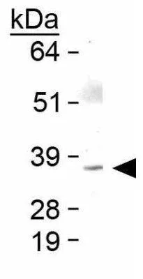
WB analysis of human skeletal muscle tissue lysate using GTX48576 Bmi1 antibody.

WB analysis of human skeletal muscle tissue lysate using GTX48576 Bmi1 antibody.
Bmi1 antibody
GTX48576
ApplicationsImmunoFluorescence, Western Blot, ImmunoCytoChemistry
Product group Antibodies
ReactivityBovine, Chicken, Feline, Human, Mouse, Primate, Rabbit, Rat, Zebra Fish
TargetBMI1
Overview
- SupplierGeneTex
- Product NameBmi1 antibody
- Delivery Days Customer9
- Application Supplier NoteWB: 2 microg/ml. ICC/IF: 1:50 - 1:200. *Optimal dilutions/concentrations should be determined by the researcher.Not tested in other applications.
- ApplicationsImmunoFluorescence, Western Blot, ImmunoCytoChemistry
- CertificationResearch Use Only
- ClonalityPolyclonal
- Concentration1 mg/ml
- ConjugateUnconjugated
- Gene ID648
- Target nameBMI1
- Target descriptionBMI1 proto-oncogene, polycomb ring finger
- Target synonymsFLVI2/BMI1, PCGF4, RNF51, flvi-2/bmi-1, polycomb complex protein BMI-1, B lymphoma Mo-MLV insertion region 1 homolog, BMI1 polycomb ring finger oncogene, BMI1 polycomb ring finger proto-oncogene, murine leukemia viral (bmi-1) oncogene homolog, polycomb group RING finger protein 4, polycomb group protein Bmi1, ring finger protein 51
- HostRabbit
- IsotypeIgG
- Protein IDP35226
- Protein NamePolycomb complex protein BMI-1
- Scientific DescriptionComponent of the Polycomb group (PcG) multiprotein PRC1 complex, a complex required to maintain the transcriptionally repressive state of many genes, including Hox genes, throughout development. PcG PRC1 complex acts via chromatin remodeling and modification of histones; it mediates monoubiquitination of histone H2A Lys-119, rendering chromatin heritably changed in its expressibility. In the PRC1 complex, it is required to stimulate the E3 ubiquitin-protein ligase activity of RNF2/RING2.
- ReactivityBovine, Chicken, Feline, Human, Mouse, Primate, Rabbit, Rat, Zebra Fish
- Storage Instruction-20°C or -80°C,2°C to 8°C
- UNSPSC41116161
Datasheet
Related products

Product group Antibodies
Anti-Bmi1 AntibodyA85203
ApplicationsWestern Blot, ELISA, ImmunoHistoChemistry
ReactivityHuman
- SizePrice
Product group Antibodies

BMI1 / PCGF4 AntibodyLS-C831530
ApplicationsWestern Blot
ReactivityMouse
TargetBMI1
- SizePrice

Product group Antibodies
Anti-BMI1 AntibodyHPA030471
ApplicationsImmunoCytoChemistry
ReactivityHuman
TargetBMI1
- SizePrice
Product group Antibodies
BMI1 AntibodyCSB-PA03345A0RB
ApplicationsWestern Blot, ELISA, ImmunoHistoChemistry
ReactivityHuman
TargetBMI1
- SizePrice
Product group Antibodies

Goat anti-BMI1 (aa237-251)EB12495
ApplicationsWestern Blot, ELISA, ImmunoHistoChemistry
ReactivityCanine, Human, Mouse, Porcine
TargetBMI1
- SizePrice
Product group Antibodies

Mouse anti Human BMI1MUB2004P
ApplicationsImmunoPrecipitation, Western Blot, ImmunoHistoChemistry, ImmunoHistoChemistry Frozen
ReactivityHuman, Mouse, Rabbit, Rat
TargetBMI1
- SizePrice
Product group Antibodies
Bmi1 antibodyGTX31296
ApplicationsImmunoFluorescence, Western Blot, ELISA, ImmunoCytoChemistry
ReactivityHuman, Mouse, Rat
TargetBMI1
- SizePrice
Product group Antibodies
BMI1 Polyclonal AntibodyCAC14038
ApplicationsWestern Blot, ELISA, ImmunoHistoChemistry
TargetBMI1
- SizePrice

Product group Antibodies
Anti-Bmi1 Antibody Picoband(r)PB9133-CARRIER-FREE
ApplicationsWestern Blot
ReactivityBovine, Human, Mouse, Rat
TargetBMI1
- SizePrice