

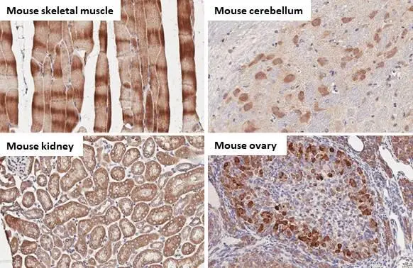
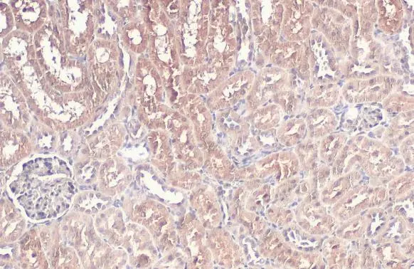
BNIP3L antibody [HL1203] detects BNIP3L protein by immunohistochemical analysis. Sample: Paraffin-embedded mouse tissues. BNIP3L stained by BNIP3L antibody [HL1203] (GTX636515) diluted at 1:100. Antigen Retrieval: Citrate buffer, pH 6.0, 15 min
![BNIP3L antibody [HL1203] detects BNIP3L protein by immunohistochemical analysis. Sample: Paraffin-embedded rat tissues. BNIP3L stained by BNIP3L antibody [HL1203] (GTX636515) diluted at 1:100. Antigen Retrieval: Citrate buffer, pH 6.0, 15 min](https://www.genetex.com/upload/website/prouct_img/normal/GTX636515/GTX636515_44501_20221230_IHC-P_multiple_R_22122821_684.webp "BNIP3L antibody [HL1203] detects BNIP3L protein by immunohistochemical analysis. Sample: Paraffin-embedded rat tissues. BNIP3L stained by BNIP3L antibody [HL1203] (GTX636515) diluted at 1:100. Antigen Retrieval: Citrate buffer, pH 6.0, 15 min")
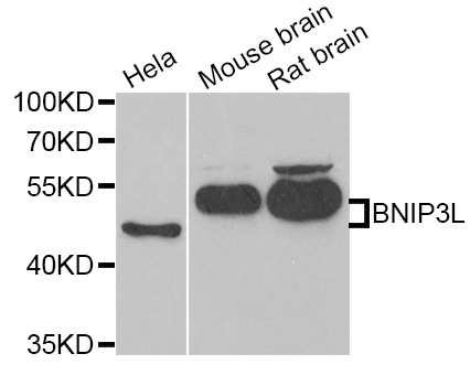
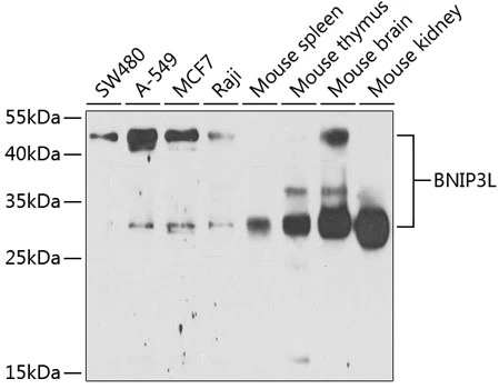
![Whole cell extract (30 μg) was separated by 12% SDS-PAGE, and the membrane was blotted with BNIP3L antibody [HL1203] (GTX636515) diluted at 1:1000. The HRP-conjugated anti-rabbit IgG antibody (GTX213110-01) was used to detect the primary antibody.](https://www.genetex.com/upload/website/prouct_img/normal/GTX636515/GTX636515_44501_20220211_WB_R_w_23061202_137.webp "Whole cell extract (30 μg) was separated by 12% SDS-PAGE, and the membrane was blotted with BNIP3L antibody [HL1203] (GTX636515) diluted at 1:1000. The HRP-conjugated anti-rabbit IgG antibody (GTX213110-01) was used to detect the primary antibody.")
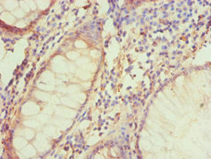
![BNIP3L antibody [HL1203] detects BNIP3L protein at mitochondria by immunohistochemical analysis. Sample: Paraffin-embedded mouse kidney. BNIP3L stained by BNIP3L antibody [HL1203] (GTX636515) diluted at 1:100. Antigen Retrieval: Citrate buffer, pH 6.0, 15 min](https://www.genetex.com/upload/website/prouct_img/normal/GTX636515/GTX636515_44501_20220318_IHC-P_M_w_23061202_197.webp "BNIP3L antibody [HL1203] detects BNIP3L protein at mitochondria by immunohistochemical analysis. Sample: Paraffin-embedded mouse kidney. BNIP3L stained by BNIP3L antibody [HL1203] (GTX636515) diluted at 1:100. Antigen Retrieval: Citrate buffer, pH 6.0, 15 min")
![BNIP3L antibody [HL1203] detects BNIP3L protein at mitochondria by immunofluorescent analysis. Sample: Mock and treated HeLa cells were fixed in 4% paraformaldehyde at RT for 15 min. Green: BNIP3L stained by BNIP3L antibody [HL1203] (GTX636515) diluted at 1:500. Blue: Fluoroshield with DAPI (GTX30920).](https://www.genetex.com/upload/website/prouct_img/normal/GTX636515/GTX636515_44501_20220318_ICC_IF_treatment_CoCl2_w_23061202_154.webp "BNIP3L antibody [HL1203] detects BNIP3L protein at mitochondria by immunofluorescent analysis. Sample: Mock and treated HeLa cells were fixed in 4% paraformaldehyde at RT for 15 min. Green: BNIP3L stained by BNIP3L antibody [HL1203] (GTX636515) diluted at 1:500. Blue: Fluoroshield with DAPI (GTX30920).")
![BNIP3L antibody [HL1203] detects BNIP3L protein at mitochondria by immunofluorescent analysis. Sample: HeLa cells were fixed in 4% paraformaldehyde at RT for 15 min. Green: BNIP3L stained by BNIP3L antibody [HL1203] (GTX636515) diluted at 1:500. Blue: Fluoroshield with DAPI (GTX30920).](https://www.genetex.com/upload/website/prouct_img/normal/GTX636515/GTX636515_44501_20220128_ICC_IF_w_23061202_550.webp "BNIP3L antibody [HL1203] detects BNIP3L protein at mitochondria by immunofluorescent analysis. Sample: HeLa cells were fixed in 4% paraformaldehyde at RT for 15 min. Green: BNIP3L stained by BNIP3L antibody [HL1203] (GTX636515) diluted at 1:500. Blue: Fluoroshield with DAPI (GTX30920).")
![Untreated (–) and treated (+) HeLa whole cell extracts (30 μg) were separated by 12% SDS-PAGE, and the membrane was blotted with BNIP3L antibody [HL1203] (GTX636515) diluted at 1:1000. The HRP-conjugated anti-rabbit IgG antibody (GTX213110-01) was used to detect the primary antibody.](https://www.genetex.com/upload/website/prouct_img/normal/GTX636515/GTX636515_44501_20211119_WB_treatment_CoCl2_w_23061202_892.webp "Untreated (–) and treated (+) HeLa whole cell extracts (30 μg) were separated by 12% SDS-PAGE, and the membrane was blotted with BNIP3L antibody [HL1203] (GTX636515) diluted at 1:1000. The HRP-conjugated anti-rabbit IgG antibody (GTX213110-01) was used to detect the primary antibody.")
BNIP3L antibody [HL1203] detects BNIP3L protein by immunohistochemical analysis. Sample: Paraffin-embedded mouse tissues. BNIP3L stained by BNIP3L antibody [HL1203] (GTX636515) diluted at 1:100. Antigen Retrieval: Citrate buffer, pH 6.0, 15 min
BNIP3L antibody [HL1203]
GTX636515
ApplicationsImmunoFluorescence, Western Blot, ImmunoCytoChemistry, ImmunoHistoChemistry, ImmunoHistoChemistry Paraffin
Product group Antibodies
ReactivityHuman, Mouse, Rat
TargetBNIP3L
Overview
- SupplierGeneTex
- Product NameBNIP3L antibody [HL1203]
- Delivery Days Customer9
- Application Supplier NoteWB: 1:500-1:3000. *Optimal dilutions/concentrations should be determined by the researcher.Not tested in other applications.
- ApplicationsImmunoFluorescence, Western Blot, ImmunoCytoChemistry, ImmunoHistoChemistry, ImmunoHistoChemistry Paraffin
- CertificationResearch Use Only
- ClonalityMonoclonal
- Clone IDHL1203
- Concentration1 mg/ml
- ConjugateUnconjugated
- Gene ID665
- Target nameBNIP3L
- Target descriptionBCL2 interacting protein 3 like
- Target synonymsBNIP3a, NIP3L, NIX, BCL2/adenovirus E1B 19 kDa protein-interacting protein 3-like, BCL2/adenovirus E1B 19 kDa protein-interacting protein 3A, BCL2/adenovirus E1B 19-kd protein-interacting protein 3a, BCL2/adenovirus E1B 19kDa interacting protein 3 like, NIP-3-like protein X, NIP3-like protein X, adenovirus E1B19k-binding protein B5
- HostRabbit
- IsotypeIgG
- Protein IDO60238
- Protein NameBCL2/adenovirus E1B 19 kDa protein-interacting protein 3-like
- Scientific DescriptionThis gene encodes a protein that belongs to the pro-apoptotic subfamily within the Bcl-2 family of proteins. The encoded protein binds to Bcl-2 and possesses the BH3 domain. The protein directly targets mitochondria and causes apoptotic changes, including loss of membrane potential and the release of cytochrome c. [provided by RefSeq, Feb 2015]
- ReactivityHuman, Mouse, Rat
- Storage Instruction-20°C or -80°C,2°C to 8°C
- UNSPSC41116161
Datasheet
Related products
Product group Antibodies
Anti-BNIP3L AntibodyA31266
ApplicationsWestern Blot, ImmunoHistoChemistry
ReactivityHuman, Mouse, Rat
- SizePrice

Product group Antibodies
Anti-BNIP3L Antibody Picoband(r)A03107-3-CARRIER-FREE
ApplicationsFlow Cytometry, ImmunoFluorescence, Western Blot, ImmunoCytoChemistry
ReactivityHuman, Mouse, Rat
TargetBNIP3L
- SizePrice
Product group Antibodies
Anti-BNIP3L Antibody144-06283
ApplicationsWestern Blot, ImmunoHistoChemistry
ReactivityHuman, Mouse, Rat
TargetBNIP3L
- SizePrice
Product group Antibodies
BNIP3L Recombinant Antibody, AbBy Fluor-594 ConjugatedBSM-61515R-BF594
ApplicationsImmunoFluorescence
ReactivityHuman
TargetBNIP3L
- SizePrice
Product group Antibodies
BNIP3L AntibodyCSB-PA002767ESR2HU
ApplicationsELISA, ImmunoHistoChemistry
ReactivityHuman
TargetBNIP3L
- SizePrice
Product group Antibodies
BNIP3L antibodyGTX28399
ApplicationsWestern Blot, ELISA, ImmunoHistoChemistry, ImmunoHistoChemistry Paraffin
ReactivityHuman
TargetBNIP3L
- SizePrice
Product group Antibodies
BNIP3L antibodyGTX111876
ApplicationsImmunoFluorescence, Western Blot, ImmunoCytoChemistry, ImmunoHistoChemistry, ImmunoHistoChemistry Paraffin
ReactivityBovine, Human, Mouse, Rat
TargetBNIP3L
- SizePrice
Product group Antibodies
BNIP3L antibodyGTX64447
ApplicationsWestern Blot, ImmunoHistoChemistry, ImmunoHistoChemistry Paraffin
ReactivityHuman, Mouse, Rat
TargetBNIP3L
- SizePrice

Product group Antibodies
Anti-BNIP3L AntibodyHPA015652
ApplicationsImmunoCytoChemistry, ImmunoHistoChemistry
ReactivityHuman
TargetBNIP3L
- SizePrice