

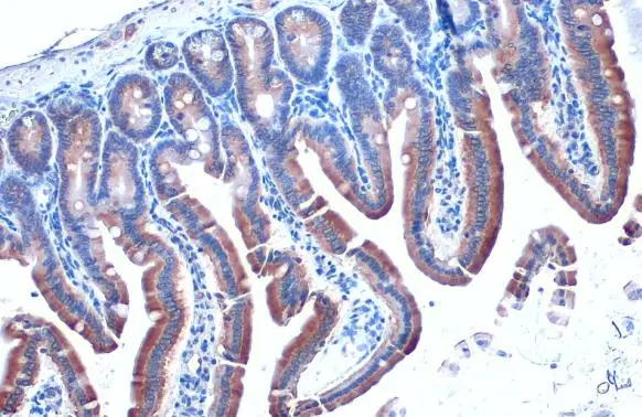
Calnexin antibody [HL1598] detects Calnexin protein at cytoplasm by immunohistochemical analysis. Sample: Paraffin-embedded mouse intestine. Calnexin stained by Calnexin antibody [HL1598] (GTX637077) diluted at 1:100. Antigen Retrieval: Citrate buffer, pH 6.0, 15 min
![Calnexin antibody [HL1598] detects Calnexin protein at endoplasmic reticulum by immunohistochemical analysis. Sample: Paraffin-embedded rat duodenum. Calnexin stained by Calnexin antibody [HL1598] (GTX637077) diluted at 1:100. Antigen Retrieval: Citrate buffer, pH 6.0, 15 min](https://www.genetex.com/upload/website/prouct_img/normal/GTX637077/GTX637077_T-44725_20220722_IHC-P_R_22080119_498.webp "Calnexin antibody [HL1598] detects Calnexin protein at endoplasmic reticulum by immunohistochemical analysis. Sample: Paraffin-embedded rat duodenum. Calnexin stained by Calnexin antibody [HL1598] (GTX637077) diluted at 1:100. Antigen Retrieval: Citrate buffer, pH 6.0, 15 min")
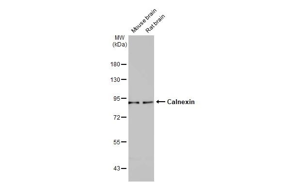
![Various tissue extracts (50 μg) were separated by 7.5% SDS-PAGE, and the membrane was blotted with Calnexin antibody [HL1598] (GTX637077) diluted at 1:1000. The HRP-conjugated anti-rabbit IgG antibody (GTX213110-01) was used to detect the primary antibody.](https://www.genetex.com/upload/website/prouct_img/normal/GTX637077/GTX637077_44788_20220902_WB_M_R_22092119_109.webp "Various tissue extracts (50 μg) were separated by 7.5% SDS-PAGE, and the membrane was blotted with Calnexin antibody [HL1598] (GTX637077) diluted at 1:1000. The HRP-conjugated anti-rabbit IgG antibody (GTX213110-01) was used to detect the primary antibody.")
![Various whole cell extracts (30 μg) were separated by 7.5% SDS-PAGE, and the membrane was blotted with Calnexin antibody [HL1598] (GTX637077) diluted at 1:1000. The HRP-conjugated anti-rabbit IgG antibody (GTX213110-01) was used to detect the primary antibody.](https://www.genetex.com/upload/website/prouct_img/normal/GTX637077/GTX637077_44788_20221209_WB_D_C_22121123_323.webp "Various whole cell extracts (30 μg) were separated by 7.5% SDS-PAGE, and the membrane was blotted with Calnexin antibody [HL1598] (GTX637077) diluted at 1:1000. The HRP-conjugated anti-rabbit IgG antibody (GTX213110-01) was used to detect the primary antibody.")
![Calnexin antibody [HL1598] detects Calnexin protein by immunohistochemical analysis. Sample: Paraffin-embedded rat tissues. Calnexin stained by Calnexin antibody [HL1598] (GTX637077) diluted at 1:100. Antigen Retrieval: Citrate buffer, pH 6.0, 15 min](https://www.genetex.com/upload/website/prouct_img/normal/GTX637077/GTX637077_44788_20221230_IHC-P_multiple_R_22122821_698.webp "Calnexin antibody [HL1598] detects Calnexin protein by immunohistochemical analysis. Sample: Paraffin-embedded rat tissues. Calnexin stained by Calnexin antibody [HL1598] (GTX637077) diluted at 1:100. Antigen Retrieval: Citrate buffer, pH 6.0, 15 min")
![Calnexin antibody [HL1598] detects Calnexin protein by immunohistochemical analysis. Sample: Paraffin-embedded mouse tissues. Calnexin stained by Calnexin antibody [HL1598] (GTX637077) diluted at 1:100. Antigen Retrieval: Citrate buffer, pH 6.0, 15 min](https://www.genetex.com/upload/website/prouct_img/normal/GTX637077/GTX637077_44788_20221230_IHC-P_multiple_M_22122821_713.webp "Calnexin antibody [HL1598] detects Calnexin protein by immunohistochemical analysis. Sample: Paraffin-embedded mouse tissues. Calnexin stained by Calnexin antibody [HL1598] (GTX637077) diluted at 1:100. Antigen Retrieval: Citrate buffer, pH 6.0, 15 min")
![Calnexin antibody [HL1598] detects Calnexin protein at cytoplasm by immunohistochemical analysis. Sample: Paraffin-embedded dog pancreas. Calnexin stained by Calnexin antibody [HL1598] (GTX637077) diluted at 1:100. Antigen Retrieval: Citrate buffer, pH 6.0, 15 min](https://www.genetex.com/upload/website/prouct_img/normal/GTX637077/GTX637077_44788_20230203_IHC-P_Dog_23020621_322.webp "Calnexin antibody [HL1598] detects Calnexin protein at cytoplasm by immunohistochemical analysis. Sample: Paraffin-embedded dog pancreas. Calnexin stained by Calnexin antibody [HL1598] (GTX637077) diluted at 1:100. Antigen Retrieval: Citrate buffer, pH 6.0, 15 min")
![Calnexin antibody [HL1598] detects Calnexin protein at cytoplasm by immunohistochemical analysis. Sample: Paraffin-embedded cat mammary gland. Calnexin stained by Calnexin antibody [HL1598] (GTX637077) diluted at 1:100. Antigen Retrieval: Citrate buffer, pH 6.0, 15 min](https://www.genetex.com/upload/website/prouct_img/normal/GTX637077/GTX637077_44788_20230203_IHC-P_Cat_23020621_601.webp "Calnexin antibody [HL1598] detects Calnexin protein at cytoplasm by immunohistochemical analysis. Sample: Paraffin-embedded cat mammary gland. Calnexin stained by Calnexin antibody [HL1598] (GTX637077) diluted at 1:100. Antigen Retrieval: Citrate buffer, pH 6.0, 15 min")
![Non-transfected (–) and transfected (+) 293T whole cell extracts (30 μg) were separated by 7.5% SDS-PAGE, and the membrane was blotted with Calnexin antibody [HL1598] (GTX637077) diluted at 1:5000. The HRP-conjugated anti-rabbit IgG antibody (GTX213110-01) was used to detect the primary antibody.](https://www.genetex.com/upload/website/prouct_img/normal/GTX637077/GTX637077_44788_20230714_WB_B_23071822_839.webp "Non-transfected (–) and transfected (+) 293T whole cell extracts (30 μg) were separated by 7.5% SDS-PAGE, and the membrane was blotted with Calnexin antibody [HL1598] (GTX637077) diluted at 1:5000. The HRP-conjugated anti-rabbit IgG antibody (GTX213110-01) was used to detect the primary antibody.")
![Non-transfected (–) and transfected (+) 293T whole cell extracts were separated by 7.5% SDS-PAGE, and the membrane was blotted with Calnexin antibody [HL1598] (GTX637077) diluted at 1:2000. The HRP-conjugated anti-rabbit IgG antibody (GTX213110-01) was used to detect the primary antibody.](https://www.genetex.com/upload/website/prouct_img/normal/GTX637077/GTX637077_44788_20230922_WB_shRNA_watermark_23100118_725.webp "Non-transfected (–) and transfected (+) 293T whole cell extracts were separated by 7.5% SDS-PAGE, and the membrane was blotted with Calnexin antibody [HL1598] (GTX637077) diluted at 1:2000. The HRP-conjugated anti-rabbit IgG antibody (GTX213110-01) was used to detect the primary antibody.")
Calnexin antibody [HL1598] detects Calnexin protein at cytoplasm by immunohistochemical analysis. Sample: Paraffin-embedded mouse intestine. Calnexin stained by Calnexin antibody [HL1598] (GTX637077) diluted at 1:100. Antigen Retrieval: Citrate buffer, pH 6.0, 15 min
Calnexin antibody [HL1598]
GTX637077
ApplicationsWestern Blot, ImmunoHistoChemistry, ImmunoHistoChemistry Paraffin
Product group Antibodies
ReactivityCanine, Feline, Human, Mouse, Rat
TargetCANX
Overview
- SupplierGeneTex
- Product NameCalnexin antibody [HL1598]
- Delivery Days Customer9
- Application Supplier NoteWB: 1:500-1:3000. *Optimal dilutions/concentrations should be determined by the researcher.Not tested in other applications.
- ApplicationsWestern Blot, ImmunoHistoChemistry, ImmunoHistoChemistry Paraffin
- CertificationResearch Use Only
- ClonalityMonoclonal
- Clone IDHL1598
- Concentration1 mg/ml
- ConjugateUnconjugated
- Gene ID821
- Target nameCANX
- Target descriptioncalnexin
- Target synonymsCNX, IP90, P90, calnexin, epididymis secretory sperm binding protein, major histocompatibility complex class I antigen-binding protein p88
- HostRabbit
- IsotypeIgG
- Protein IDP27824
- Protein NameCalnexin
- Scientific DescriptionThis gene encodes a member of the calnexin family of molecular chaperones. The encoded protein is a calcium-binding, endoplasmic reticulum (ER)-associated protein that interacts transiently with newly synthesized N-linked glycoproteins, facilitating protein folding and assembly. It may also play a central role in the quality control of protein folding by retaining incorrectly folded protein subunits within the ER for degradation. Alternatively spliced transcript variants encoding different isoforms have been described. [provided by RefSeq, Jun 2018]
- ReactivityCanine, Feline, Human, Mouse, Rat
- Storage Instruction-20°C or -80°C,2°C to 8°C
- UNSPSC12352203
Datasheet
Related products
Product group Antibodies
Anti-CANX Antibody144-63846
ApplicationsImmunoFluorescence, Western Blot, ImmunoHistoChemistry
ReactivityHuman, Mouse, Rat
TargetCANX
- SizePrice

Product group Antibodies
Anti-Calnexin/CANX Antibody Picoband(r)A03372-2-CARRIER-FREE
ApplicationsFlow Cytometry, ImmunoFluorescence, Western Blot, ELISA, ImmunoCytoChemistry, ImmunoHistoChemistry
ReactivityHuman, Mouse
TargetCANX
- SizePrice
![Various whole cell extracts (30 μg) were separated by 7.5% SDS-PAGE, and the membrane was blotted with Calnexin antibody [N3C2], Internal (GTX101676) diluted at 1:5000. The HRP-conjugated anti-rabbit IgG antibody (GTX213110-01) was used to detect the primary antibody.](https://www.genetex.com/upload/website/prouct_img/normal/GTX101676/GTX101676_43643_20190719_WB_w_23060100_608.webp)
Product group Antibodies
References
Calnexin antibody [N3C2], InternalGTX101676
ApplicationsImmunoFluorescence, Western Blot, ImmunoCytoChemistry, ImmunoHistoChemistry, ImmunoHistoChemistry Paraffin
ReactivityHuman, Mouse, Rat
TargetCANX
- SizePrice
![Calnexin antibody [C3], C-term detects Calnexin protein at cell membrane and cytoplasm by immunohistochemical analysis. Sample: Paraffin-embedded rat brain. Calnexin stained by Calnexin antibody [C3], C-term (GTX109669) diluted at 1:165. Antigen Retrieval: Citrate buffer, pH 6.0, 15 min](https://www.genetex.com/upload/website/prouct_img/normal/GTX109669/GTX109669_44699_20220701_IHC-P_R_22071401_447.webp)
Product group Antibodies
References
Calnexin antibody [C3], C-termGTX109669
ApplicationsImmunoFluorescence, ImmunoPrecipitation, Western Blot, ImmunoCytoChemistry, ImmunoHistoChemistry, ImmunoHistoChemistry Paraffin
ReactivityHuman, Mouse, Rat, Sheep
TargetCANX
- SizePrice
Product group Antibodies
References
Calnexin antibodyGTX112886
ApplicationsImmunoFluorescence, Western Blot, ImmunoCytoChemistry, ImmunoHistoChemistry, ImmunoHistoChemistry Paraffin
ReactivityHuman, Mouse, Rat
TargetCANX
- SizePrice
Product group Antibodies
Calnexin antibody [AT18B9]GTX57717
ApplicationsFlow Cytometry, ImmunoFluorescence, Western Blot, ImmunoCytoChemistry
ReactivityHuman
TargetCANX
- SizePrice
![Calnexin antibody [GT1563] detects Calnexin protein at cytoplasm by immunofluorescent analysis. Sample: Neuro2A cells were fixed in 4% paraformaldehyde at RT for 15 min. Green: Calnexin stained by Calnexin antibody [GT1563] (GTX629976) diluted at 1:500. Blue: Fluoroshield with DAPI (GTX30920).](https://www.genetex.com/upload/website/prouct_img/normal/GTX629976/GTX629976_41484_20230512_ICC_IF_M_23060622_501.webp)
Product group Antibodies
References
Calnexin antibody [GT1563]GTX629976
ApplicationsDot Blot, ImmunoFluorescence, ImmunoPrecipitation, Western Blot, ImmunoCytoChemistry
ReactivityHuman, Mouse, Rat
TargetCANX
- SizePrice
Product group Antibodies
CANX Polyclonal AntibodyCAC14640
ApplicationsImmunoFluorescence, Western Blot, ELISA, ImmunoHistoChemistry
TargetCANX
- SizePrice
Product group Antibodies
References
Calnexin Polyclonal AntibodyBS-1693R
ApplicationsFlow Cytometry, ImmunoFluorescence, Western Blot, ELISA, ImmunoCytoChemistry, ImmunoHistoChemistry, ImmunoHistoChemistry Frozen, ImmunoHistoChemistry Paraffin
ReactivityBovine, Chicken, Equine, Human, Mouse, Rabbit, Rat
TargetCANX
- SizePrice
Product group Antibodies
CANX AntibodyCSB-PA001175
ApplicationsImmunoFluorescence, Western Blot, ELISA, ImmunoHistoChemistry
ReactivityHuman, Mouse, Rat
TargetCANX
- SizePrice