

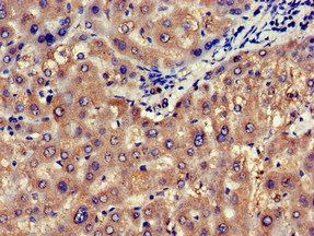
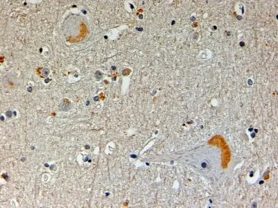
CAPON Polyclonal Antibody
BS-11987R
ApplicationsImmunoFluorescence, Western Blot, ELISA, ImmunoCytoChemistry, ImmunoHistoChemistry, ImmunoHistoChemistry Frozen, ImmunoHistoChemistry Paraffin
Product group Antibodies
ReactivityBovine, Equine, Human, Mouse, Rat, Sheep
Overview
- SupplierBioss
- Product NameCAPON Polyclonal Antibody
- Delivery Days Customer16
- ApplicationsImmunoFluorescence, Western Blot, ELISA, ImmunoCytoChemistry, ImmunoHistoChemistry, ImmunoHistoChemistry Frozen, ImmunoHistoChemistry Paraffin
- Applications SupplierWB(1:300-5000), ELISA(1:500-1000), IHC-P(1:200-400), IHC-F(1:100-500), IF(IHC-P)(1:50-200), IF(IHC-F)(1:50-200), IF(ICC)(1:50-200)
- CertificationResearch Use Only
- ClonalityPolyclonal
- Concentration1 ug/ul
- ConjugateUnconjugated
- HostRabbit
- IsotypeIgG
- ReactivityBovine, Equine, Human, Mouse, Rat, Sheep
- Storage Instruction-20°C
- UNSPSC41116161
Datasheet
Related products

Product group Antibodies
Anti-CAPON/NOS1AP Antibody Picoband(r)A03060-3-CARRIER-FREE
ApplicationsFlow Cytometry, Western Blot, ELISA, ImmunoHistoChemistry
ReactivityHuman, Mouse, Rat
TargetNOS1AP
- SizePrice
Product group Antibodies

NOS1AP / CAPON AntibodyLS-C678837
ApplicationsImmunoFluorescence, ELISA, ImmunoHistoChemistry, ImmunoHistoChemistry Paraffin
ReactivityHuman
TargetNOS1AP
- SizePrice
Product group Antibodies

Goat anti-CAPON / NOS1APEB05583
ApplicationsELISA, ImmunoHistoChemistry
ReactivityBovine, Human, Mouse, Rat
TargetNOS1AP
- SizePrice
Product group Antibodies
ApplicationsImmunoPrecipitation, Western Blot, ImmunoCytoChemistry, ImmunoHistoChemistry
ReactivityMouse, Rat
TargetNOS1AP
- SizePrice
Product group Antibodies
NOS1AP AntibodyCSB-PA015942LA01HU
ApplicationsImmunoFluorescence, ELISA, ImmunoHistoChemistry
ReactivityHuman
TargetNOS1AP
- SizePrice
Product group Antibodies
CAPON antibody, N-termGTX89878
ApplicationsImmunoHistoChemistry, ImmunoHistoChemistry Paraffin
ReactivityHuman
TargetNOS1AP
- SizePrice

Product group Antibodies
Anti-NOS1AP AntibodyHPA030066
ApplicationsWestern Blot, ImmunoCytoChemistry, ImmunoHistoChemistry
ReactivityHuman
TargetNOS1AP
- SizePrice
Product group Antibodies
NOS1AP AntibodyPACO51750
ApplicationsImmunoFluorescence, ELISA, ImmunoHistoChemistry
ReactivityHuman
TargetNOS1AP
- SizePrice