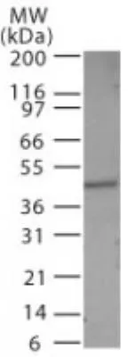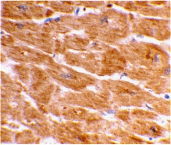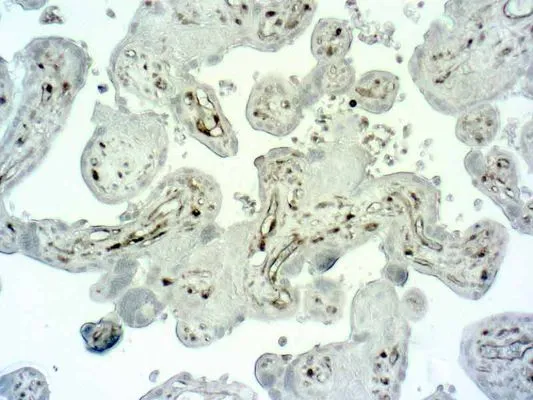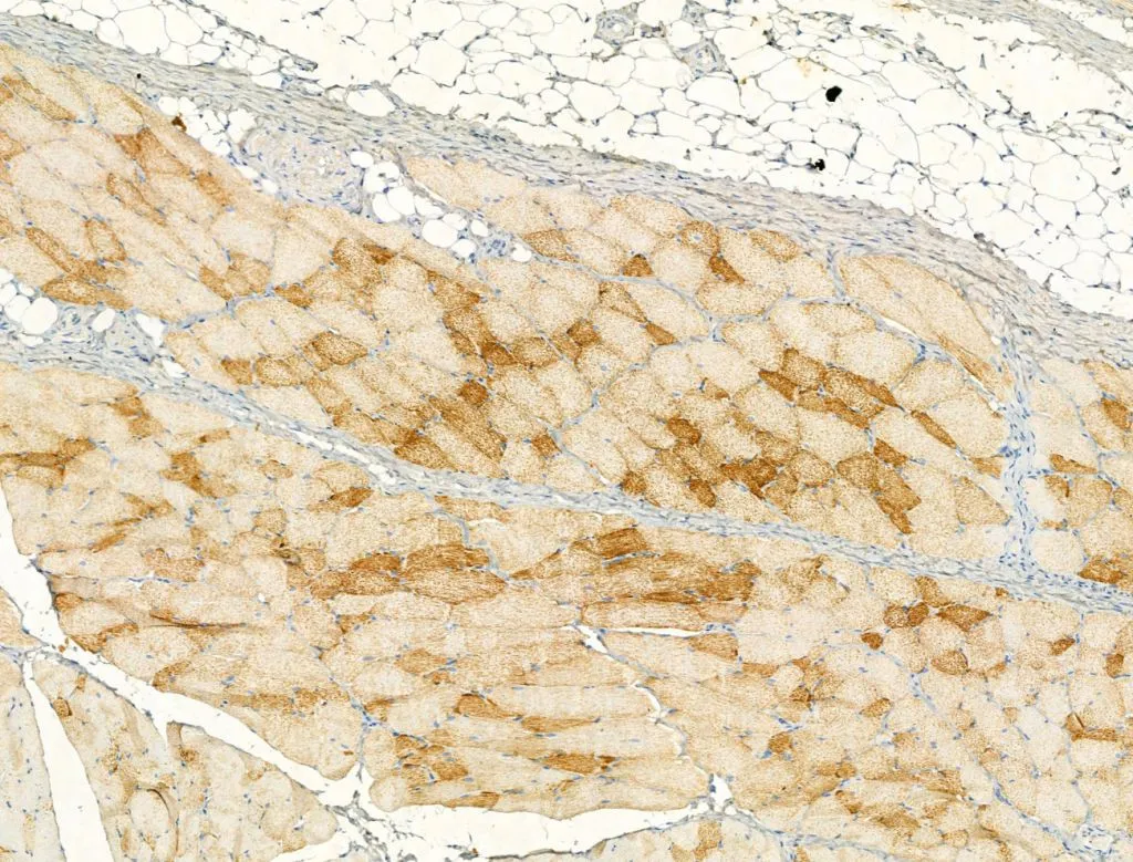
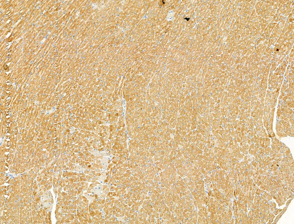
IHC-P analysis of rat skin tissue using GTX03738 Caspase 1 (cleaved Asp297) antibody. Antigen retrieval : Heat mediated antigen retrieval step in citrate buffer was performed Dilution : 1:100
Caspase 1 (cleaved Asp297) antibody
GTX03738
ApplicationsImmunoFluorescence, Western Blot, ImmunoCytoChemistry, ImmunoHistoChemistry, ImmunoHistoChemistry Paraffin
Product group Antibodies
TargetCASP1
Overview
- SupplierGeneTex
- Product NameCaspase 1 (cleaved Asp297) antibody
- Delivery Days Customer9
- Application Supplier NoteWB: 1:500-1:2000. IHC-P: 1:50-1:200. *Optimal dilutions/concentrations should be determined by the researcher.Not tested in other applications.
- ApplicationsImmunoFluorescence, Western Blot, ImmunoCytoChemistry, ImmunoHistoChemistry, ImmunoHistoChemistry Paraffin
- CertificationResearch Use Only
- ClonalityPolyclonal
- Concentration1 mg/ml
- ConjugateUnconjugated
- Gene ID834
- Target nameCASP1
- Target descriptioncaspase 1
- Target synonymsCASP1 nirs variant 1; caspase 1, apoptosis-related cysteine peptidase; caspase-1; caspase-1 isoform alpha; ICE; IL-1 beta-converting enzyme; IL1BC; IL1B-convertase; interleukin 1, beta, convertase; interleukin 1-B converting enzyme; P45
- HostRabbit
- IsotypeIgG
- Protein IDP29466
- Protein NameCaspase-1
- Scientific DescriptionThis gene encodes a protein which is a member of the cysteine-aspartic acid protease (caspase) family. Sequential activation of caspases plays a central role in the execution-phase of cell apoptosis. Caspases exist as inactive proenzymes which undergo proteolytic processing at conserved aspartic residues to produce 2 subunits, large and small, that dimerize to form the active enzyme. This gene was identified by its ability to proteolytically cleave and activate the inactive precursor of interleukin-1, a cytokine involved in the processes such as inflammation, septic shock, and wound healing. This gene has been shown to induce cell apoptosis and may function in various developmental stages. Studies of a similar gene in mouse suggest a role in the pathogenesis of Huntington disease. Alternative splicing results in transcript variants encoding distinct isoforms. [provided by RefSeq, Mar 2012]
- Storage Instruction-20°C or -80°C,2°C to 8°C
- UNSPSC12352203

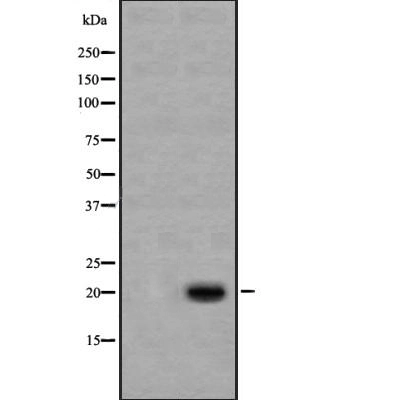
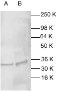
![WB analysis of HeLa (A) and NIH-3T3 (B) cell lysate using GTX14367 Caspase 1 antibody [14F468]. Dilution : 0.5 microg/ml and 2 microg/ml](https://www.genetex.com/upload/website/prouct_img/normal/GTX14367/GTX14367_1077_WB_w_23060620_226.webp)
