


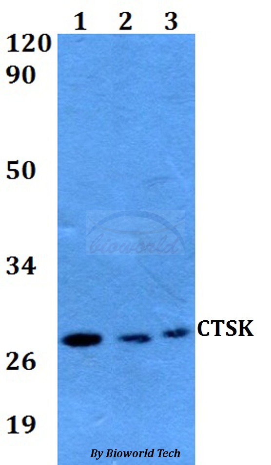
WB analysis of 293 cell lysate (2 ug/lane) either nontransfected (Lane 1) or transiently transfected with the Cathepsin K (Lane 2) using GTX81488 Cathepsin K antibody, Internal.


WB analysis of 293 cell lysate (2 ug/lane) either nontransfected (Lane 1) or transiently transfected with the Cathepsin K (Lane 2) using GTX81488 Cathepsin K antibody, Internal.
Cathepsin K antibody, Internal
GTX81488
ApplicationsFlow Cytometry, Western Blot, ImmunoHistoChemistry, ImmunoHistoChemistry Paraffin
Product group Antibodies
ReactivityHuman
TargetCTSK
Overview
- SupplierGeneTex
- Product NameCathepsin K antibody, Internal
- Delivery Days Customer9
- Application Supplier NoteWB: 1:1000. IHC-P: 1:10-1:50. FCM: 1:10-1:50. *Optimal dilutions/concentrations should be determined by the researcher.Not tested in other applications.
- ApplicationsFlow Cytometry, Western Blot, ImmunoHistoChemistry, ImmunoHistoChemistry Paraffin
- CertificationResearch Use Only
- ClonalityPolyclonal
- ConjugateUnconjugated
- Gene ID1513
- Target nameCTSK
- Target descriptioncathepsin K
- Target synonymsCTS02, CTSO, CTSO1, CTSO2, PKND, PYCD, cathepsin K, cathepsin O, cathepsin O1, cathepsin O2, cathepsin X
- HostRabbit
- IsotypeIgG
- Protein IDP43235
- Protein NameCathepsin K
- Scientific DescriptionThe protein encoded by this gene is a lysosomal cysteine proteinase involved in bone remodeling and resorption. This protein, which is a member of the peptidase C1 protein family, is predominantly expressed in osteoclasts. However, the encoded protein is also expressed in a significant fraction of human breast cancers, where it could contribute to tumor invasiveness. Mutations in this gene are the cause of pycnodysostosis, an autosomal recessive disease characterized by osteosclerosis and short stature. [provided by RefSeq, Apr 2013]
- ReactivityHuman
- Storage Instruction-20°C or -80°C,2°C to 8°C
- UNSPSC41116161
Datasheet
Related products
Product group Antibodies
Anti-CTSK AntibodyA28771
ApplicationsWestern Blot
ReactivityHuman, Mouse, Rat
- SizePrice
Product group Antibodies
References
Cathepsin K Polyclonal AntibodyBS-1611R
ApplicationsImmunoFluorescence, Western Blot, ImmunoHistoChemistry, ImmunoHistoChemistry Frozen, ImmunoHistoChemistry Paraffin
ReactivityMouse, Rat
TargetCTSK
- SizePrice
Product group Antibodies
CTSK AntibodyCSB-PA006192ESR1HU
ApplicationsWestern Blot, ELISA, ImmunoHistoChemistry
ReactivityHuman, Rat
TargetCTSK
- SizePrice
Product group Antibodies

Goat anti-Cathepsin KEB08596
ApplicationsImmunoFluorescence, Western Blot, ELISA
ReactivityBovine, Canine, Human, Mouse, Porcine, Rat
TargetCTSK
- SizePrice
Product group Antibodies
Ctsk Polyclonal AntibodyCAC10607
ApplicationsWestern Blot, ELISA, ImmunoHistoChemistry
ReactivityRat
TargetCTSK
- SizePrice
Product group Antibodies
ApplicationsWestern Blot, ELISA
ReactivityHuman
TargetCTSK
- SizePrice

Product group Antibodies
Anti-CTSK AntibodyHPA050346
ApplicationsImmunoCytoChemistry
ReactivityHuman
TargetCTSK
- SizePrice

Product group Antibodies
Anti-Cathepsin K/CTSK Antibody Picoband(r)PB9856-CARRIER-FREE
ApplicationsWestern Blot, ImmunoHistoChemistry
ReactivityHuman
TargetCTSK
- SizePrice
Product group Antibodies

CTSK / Cathepsin K AntibodyLS-C331688
ApplicationsWestern Blot, ImmunoHistoChemistry
ReactivityAvian, Human, Mouse, Rat
TargetCTSK
- SizePrice