

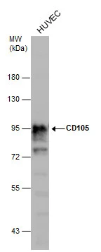
Whole cell extract (30 μg) was separated by 7.5% SDS-PAGE, and the membrane was blotted with CD105 antibody [N1N3] (GTX112684) diluted at 1:1000.
![CD105 antibody [N1N3] detects CD105 protein at cell membrane and cytoplasm by immunohistochemical analysis. Sample: Paraffin-embedded mouse brown adipocyte. CD105 stained by CD105 antibody [N1N3] (GTX112684) diluted at 1:500. Antigen Retrieval: Citrate buffer, pH 6.0, 15 min](https://www.genetex.com/upload/website/prouct_img/normal/GTX112684/GTX112684_40492_20191101_IHC-P_M_w_23060500_731.webp "CD105 antibody [N1N3] detects CD105 protein at cell membrane and cytoplasm by immunohistochemical analysis. Sample: Paraffin-embedded mouse brown adipocyte. CD105 stained by CD105 antibody [N1N3] (GTX112684) diluted at 1:500. Antigen Retrieval: Citrate buffer, pH 6.0, 15 min")
Whole cell extract (30 μg) was separated by 7.5% SDS-PAGE, and the membrane was blotted with CD105 antibody [N1N3] (GTX112684) diluted at 1:1000.
CD105 antibody [N1N3]
GTX112684
ApplicationsWestern Blot, ImmunoHistoChemistry, ImmunoHistoChemistry Paraffin
Product group Antibodies
ReactivityHuman, Mouse
TargetENG
Overview
- SupplierGeneTex
- Product NameCD105 antibody [N1N3]
- Delivery Days Customer9
- Application Supplier NoteWB: 1:1000-1:10000. *Optimal dilutions/concentrations should be determined by the researcher.Not tested in other applications.
- ApplicationsWestern Blot, ImmunoHistoChemistry, ImmunoHistoChemistry Paraffin
- CertificationResearch Use Only
- ClonalityPolyclonal
- Concentration1 mg/ml
- ConjugateUnconjugated
- Gene ID2022
- Target nameENG
- Target descriptionendoglin
- Target synonymsEND, HHT1, ORW1, endoglin, CD105 antigen, soluble endoglin
- HostRabbit
- IsotypeIgG
- Protein IDP17813
- Protein NameEndoglin
- Scientific DescriptionThis gene encodes a homodimeric transmembrane protein which is a major glycoprotein of the vascular endothelium. This protein is a component of the transforming growth factor beta receptor complex and it binds TGFB1 and TGFB3 with high affinity. Mutations in this gene cause hereditary hemorrhagic telangiectasia, also known as Osler-Rendu-Weber syndrome 1, an autosomal dominant multisystemic vascular dysplasia. Alternatively spliced transcript variants encoding different isoforms have been found for this gene. [provided by RefSeq]
- ReactivityHuman, Mouse
- Storage Instruction-20°C or -80°C,2°C to 8°C
- UNSPSC12352203
Datasheet
Related products
Product group Antibodies

Anti-CD105 [huRH105-1]AB04017-1.1
ApplicationsFlow Cytometry, Other Application
ReactivityHuman, Rat
TargetENG
- SizePrice
Product group Antibodies
Anti-Endoglin Antibody130-10018
ApplicationsELISA
ReactivityHuman
TargetENG
- SizePrice

Product group Antibodies
Anti-CD105/ENG Antibody Picoband(r)A02997-1-CARRIER-FREE
ApplicationsFlow Cytometry, Western Blot
ReactivityHuman
TargetENG
- SizePrice
![FCM analysis of mouse bone marrow cells using GTX00572-08 CD105 antibody [MJ7/18] (PE).](https://www.genetex.com/upload/website/prouct_img/normal/GTX00572-08/GTX00572-08_20250623_FCM_25062300_103.webp)
Product group Antibodies
References
CD105 antibody [MJ7/18] (PE)GTX00572-08
ApplicationsFlow Cytometry
ReactivityHuman, Mouse, Rat
TargetENG
- SizePrice
![Whole cell extract (30 μg) was separated by 7.5% SDS-PAGE, and the membrane was blotted with CD105 antibody [N3C3] (GTX100508) diluted at 1:1000.](https://www.genetex.com/upload/website/prouct_img/normal/GTX100508/GTX100508_39932_20151119_WB_w_23060100_822.webp)
Product group Antibodies
CD105 antibody [N3C3]GTX100508
ApplicationsWestern Blot, ImmunoHistoChemistry, ImmunoHistoChemistry Paraffin
ReactivityHuman, Mouse
TargetENG
- SizePrice
![CD105 antibody [N1N3-2] detects CD105 protein at cell membrane and cytoplasm by immunohistochemical analysis. Sample: Paraffin-embedded human esophageal carcinoma. CD105 stained by CD105 antibody [N1N3-2] (GTX112685) diluted at 1:500. Antigen Retrieval: Citrate buffer, pH 6.0, 15 min](https://www.genetex.com/upload/website/prouct_img/normal/GTX112685/GTX112685_40499_20191115_IHC-P_w_23060500_674.webp)
Product group Antibodies
CD105 antibody [N1N3-2]GTX112685
ApplicationsWestern Blot, ImmunoHistoChemistry, ImmunoHistoChemistry Paraffin
ReactivityHuman, Mouse
TargetENG
- SizePrice
![ELISA analysis of antigen using GTX60452 CD105 antibody [3A9].
Red : Control antigen 100ng
Purple : Antigen 10ng
Green : Antigen 50ng
Blue : Antigen 100ng](https://www.genetex.com/upload/website/prouct_img/normal/GTX60452/GTX60452_20170912_ELISA_w_23061123_556.webp)
Product group Antibodies
CD105 antibody [3A9]GTX60452
ApplicationsFlow Cytometry, ImmunoFluorescence, Western Blot, ELISA, ImmunoCytoChemistry, ImmunoHistoChemistry, ImmunoHistoChemistry Paraffin
ReactivityHuman
TargetENG
- SizePrice
![FACS analysis of KG1 cells using GTX75142 CD105 antibody [SN6] (PE).](https://www.genetex.com/upload/website/prouct_img/normal/GTX75142/GTX75142_245_FACS_w_23061322_133.webp)
Product group Antibodies
References
CD105 antibody [SN6] (PE)GTX75142
ApplicationsFlow Cytometry
ReactivityEquine, Human, Monkey, Primate, Rabbit
TargetENG
- SizePrice
Product group Antibodies
ApplicationsFlow Cytometry
ReactivityHuman
TargetENG
- SizePrice