

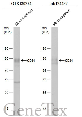
Mouse tissue extract (50 μg) was separated by 7.5% SDS-PAGE, and the membranes were blotted with CD31 antibody (GTX130274) diluted at 1:500 and competitor's antibody (ab124432) diluted at 1:500. The HRP-conjugated anti-rabbit IgG antibody (GTX213110-01) was used to detect the primary antibody.
 diluted at 1:2000.
Antigen Retrieval: Citrate buffer, pH 6.0, 15 min")
 diluted at 1:1000. Antigen Retrieval: Citrate buffer, pH 6.0, 15 min")
![CD31 antibody detects CD31 protein at cell membrane by immunohistochemical analysis. Sample: Paraffin-embedded mouse placenta. Green: CD31 stained by CD31 antibody (GTX130274) diluted at 1:250. Red: beta Actin, a Cytoskeleton marker, stained by beta Actin antibody [GT5512] (GTX629630) diluted at 1:500. Blue: Fluoroshield with DAPI (GTX30920). Antigen Retrieval: Citrate buffer, pH 6.0, 15 min](https://www.genetex.com/upload/website/prouct_img/normal/GTX130274/GTX130274_41976_20231208_IHC-P-FL_M_23121922_844.webp "CD31 antibody detects CD31 protein at cell membrane by immunohistochemical analysis. Sample: Paraffin-embedded mouse placenta. Green: CD31 stained by CD31 antibody (GTX130274) diluted at 1:250. Red: beta Actin, a Cytoskeleton marker, stained by beta Actin antibody [GT5512] (GTX629630) diluted at 1:500. Blue: Fluoroshield with DAPI (GTX30920). Antigen Retrieval: Citrate buffer, pH 6.0, 15 min")
 was separated by 7.5% SDS-PAGE, and the membrane was blotted with CD31 antibody (GTX130274) diluted at 1:500. The HRP-conjugated anti-rabbit IgG antibody (GTX213110-01) was used to detect the primary antibody.")
![CD31 antibody detects CD31 protein by immunohistochemical analysis. Sample: Frozen-sectioned mouse cerebellum. Green: CD31 stained by CD31 antibody (GTX130274) diluted at 1:250. Red: alpha Tubulin antibody [GT114] (GTX628802) diluted at 1:1000. Blue: Hoechst 33342 staining.](https://www.genetex.com/upload/website/prouct_img/normal/GTX130274/GTX130274_44909_20250627_IHC-Fr_M_25070323_684.webp "CD31 antibody detects CD31 protein by immunohistochemical analysis. Sample: Frozen-sectioned mouse cerebellum. Green: CD31 stained by CD31 antibody (GTX130274) diluted at 1:250. Red: alpha Tubulin antibody [GT114] (GTX628802) diluted at 1:1000. Blue: Hoechst 33342 staining.")
Mouse tissue extract (50 μg) was separated by 7.5% SDS-PAGE, and the membranes were blotted with CD31 antibody (GTX130274) diluted at 1:500 and competitor's antibody (ab124432) diluted at 1:500. The HRP-conjugated anti-rabbit IgG antibody (GTX213110-01) was used to detect the primary antibody.
CD31 antibody
GTX130274
ApplicationsWestern Blot, ImmunoHistoChemistry, ImmunoHistoChemistry Frozen, ImmunoHistoChemistry Paraffin
Product group Antibodies
ReactivityHuman, Mouse
TargetPecam1
Overview
- SupplierGeneTex
- Product NameCD31 antibody
- Delivery Days Customer9
- Application Supplier NoteWB: 1:500-1:3000. IHC-P: 1:100-1:1000. *Optimal dilutions/concentrations should be determined by the researcher.Not tested in other applications.
- ApplicationsWestern Blot, ImmunoHistoChemistry, ImmunoHistoChemistry Frozen, ImmunoHistoChemistry Paraffin
- CertificationResearch Use Only
- ClonalityPolyclonal
- Concentration1.44 mg/ml
- ConjugateUnconjugated
- Gene ID18613
- Target namePecam1
- Target descriptionplatelet/endothelial cell adhesion molecule 1
- Target synonymsCd31, PECAM-1, Pecam, platelet endothelial cell adhesion molecule
- HostRabbit
- IsotypeIgG
- Protein IDQ08481
- Protein NamePlatelet endothelial cell adhesion molecule
- Scientific DescriptionCell adhesion molecule which is required for leukocyte transendothelial migration (TEM) under most inflammatory conditions. Tyr-679 plays a critical role in TEM and is required for efficient trafficking of PECAM1 to and from the lateral border recycling compartment (LBRC) and is also essential for the LBRC membrane to be targeted around migrating leukocytes. Prevents phagocyte ingestion of closely apposed viable cells by transmitting detachment signals, and changes function on apoptosis, promoting tethering of dying cells to phagocytes (the encounter of a viable cell with a phagocyte via the homophilic interaction of PECAM1 on both cell surfaces leads to the viable cells active repulsion from the phagocyte. During apoptosis, the inside-out signaling of PECAM1 is somehow disabled so that the apoptotic cell does not actively reject the phagocyte anymore. The lack of this repulsion signal together with the interaction of the eat-me signals and their respective receptors causes the attachment of the apoptotic cell to the phagocyte, thus triggering the process of engulfment). Modulates BDKRB2 activation (By similarity). Induces susceptibility to atherosclerosis.
- ReactivityHuman, Mouse
- Storage Instruction-20°C or -80°C,2°C to 8°C
- UNSPSC41116161
Datasheet
Related products
Product group Antibodies

M CD31 AntibodyABX025991
ApplicationsFlow Cytometry, Western Blot, ELISA
- SizePrice

Product group Antibodies
Anti-CD31/Pecam1 Antibody Picoband(r)A01513-1-CARRIER-FREE
ApplicationsWestern Blot, ELISA, ImmunoHistoChemistry
ReactivityMouse
TargetPecam1
- SizePrice
Product group Antibodies

ApplicationsWestern Blot, ELISA
ReactivityMouse, Rat
TargetPecam1
- SizePrice
Product group Antibodies
ApplicationsImmunoPrecipitation, Western Blot, ImmunoCytoChemistry, ImmunoHistoChemistry
ReactivityMouse
TargetPecam1
- SizePrice
Product group Antibodies
CD31 antibody [2H8] (Azide free)GTX74943
ApplicationsFlow Cytometry, ImmunoFluorescence, ImmunoCytoChemistry, ImmunoHistoChemistry, Neutralisation/Blocking
ReactivityMouse
TargetPecam1
- SizePrice
![IHC-P analysis of kidney, lung, heart and liver tissue sections from LPS exposed mouse using GTX53123 CD31 antibody [1D12].](https://www.genetex.com/upload/website/prouct_img/normal/GTX53123/GTX53123_20191119_IHC-P_w_23060900_255.webp)
Product group Antibodies
References
CD31 antibody [1D12]GTX53123
ApplicationsImmunoHistoChemistry, ImmunoHistoChemistry Frozen, ImmunoHistoChemistry Paraffin
ReactivityMouse
TargetPecam1
- SizePrice
Product group Antibodies
References
CD31 antibody [MEC7.46]GTX54379
ApplicationsFlow Cytometry, ImmunoFluorescence, ImmunoPrecipitation, ImmunoCytoChemistry, ImmunoHistoChemistry, ImmunoHistoChemistry Frozen, ImmunoHistoChemistry Paraffin
ReactivityMouse
TargetPecam1
- SizePrice
Product group Antibodies
CD31 antibody [ER-MP12]GTX54402
ApplicationsFlow Cytometry, ImmunoPrecipitation, Western Blot, ELISA, ImmunoHistoChemistry, ImmunoHistoChemistry Frozen, ImmunoHistoChemistry Paraffin
ReactivityMouse
TargetPecam1
- SizePrice
Product group Antibodies
Anti-CD31/PECAM1 AntibodyCAB0378
ApplicationsImmunoFluorescence, Western Blot, ELISA, ImmunoCytoChemistry, ImmunoHistoChemistry, ImmunoHistoChemistry Paraffin
ReactivityHuman
TargetPecam1
- SizePrice