

CD33(C33/68), Biotin conjugate, 0.1mg/mL [26628-22-8]
BNCB0068
ReactivityBovine, Human, Mouse
Product group Antibodies
TargetCD33
Overview
- SupplierBiotium
- Product NameCD33(C33/68), Biotin conjugate, 0.1mg/mL [26628-22-8]
- Delivery Days Customer9
- CAS Number26628-22-8
- CertificationResearch Use Only
- ClonalityMonoclonal
- Clone IDC33/68
- Concentration0.1 mg/ml
- ConjugateBiotin
- Gene ID945
- Target nameCD33
- Target descriptionCD33 molecule
- Target synonymsCD33rSiglec, SIGLEC-3, SIGLEC3, p67, myeloid cell surface antigen CD33, CD33 antigen (gp67), CD33 molecule transcript, gp67, sialic acid-binding Ig-like lectin 3
- HostMouse
- IsotypeIgG1
- Protein IDP20138
- Protein NameMyeloid cell surface antigen CD33
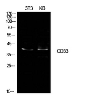
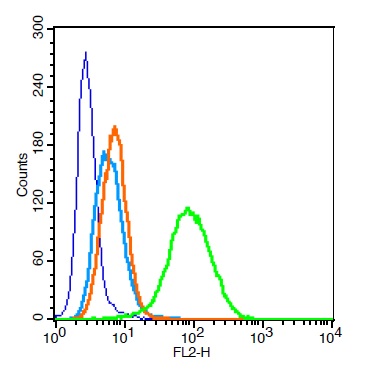
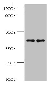
- Scientific DescriptionRecognizes a 67 kDa glycoprotein, which is identified as CD33. It is a transmembrane protein of the sialic acid-binding immunoglobulin-like lectin (Siglec) family. It belongs to the immunoreceptor tyrosine-based inhibitory motif (ITIM)-containing molecules able of recruiting protein tyrosine phosphatases SHP-1 and SHP-2 to signal assemblies; these ITIMs are also used for ubiquitin-mediated removal of the receptor from the cell surface. CD33 is expressed on cells of myelomonocytic lineage, binds sialic acid residues in N- and O-glycans on cell surfaces, and is a therapeutic target for acute myeloid leukemia. CD33 is expressed on myeloid progenitors, monocytes, granulocytes, dendritic cells and mast cells. It is absent on platelets, lymphocytes, erythrocytes and hematopoietic stem cells.Primary antibodies are available purified, or with a selection of fluorescent CF® Dyes and other labels. CF® Dyes offer exceptional brightness and photostability. Note: Conjugates of blue fluorescent dyes like CF®405S and CF®405M are not recommended for detecting low abundance targets, because blue dyes have lower fluorescence and can give higher non-specific background than other dye colors.
- SourceAnimal
- ReactivityBovine, Human, Mouse
- Storage Instruction2°C to 8°C,RT
- UNSPSC41116161
MSDS
Related products
Product group Antibodies

Anti-CD33 [hP67.6 (Gemtuzumab)]Ab00283-1.1
ApplicationsFlow Cytometry, ImmunoFluorescence
ReactivityHuman
TargetCD33
- SizePrice
Product group Antibodies
Anti-CD33 AntibodyA101517
ApplicationsWestern Blot, ELISA
ReactivityHuman
- SizePrice
Product group Antibodies
CD33 Polyclonal AntibodyBS-2042R
ApplicationsFlow Cytometry, ImmunoFluorescence, Western Blot, ELISA, ImmunoCytoChemistry, ImmunoHistoChemistry, ImmunoHistoChemistry Frozen, ImmunoHistoChemistry Paraffin
ReactivityHuman
TargetCD33
- SizePrice
Product group Antibodies
Cd33 Polyclonal AntibodyCAC11135
ApplicationsWestern Blot, ELISA, ImmunoHistoChemistry
TargetCD33
- SizePrice
Product group Antibodies
CD33 AntibodyCSB-PA004925ESR1HU
ApplicationsWestern Blot, ELISA, ImmunoHistoChemistry
ReactivityHuman
TargetCD33
- SizePrice
Product group Antibodies
CD33 antibody [WM53] (PE-Cy7)GTX00477-10
ApplicationsFlow Cytometry
ReactivityHuman, Primate
TargetCD33
- SizePrice
Product group Antibodies

MERS-CoV Nucleoprotein AntibodyLS-C488273
ApplicationsELISA
ReactivityVirus
- SizePrice