


CD4, Mouse (T-Cell Marker)(GK1.5), CF700 conjugate, 0.1mg/mL [26628-22-8]
BNC002009
ApplicationsFunctional Assay, Flow Cytometry
Product group Antibodies
ReactivityBovine, Mouse, Rat
TargetFCGR1A
Overview
- SupplierBiotium
- Product NameCD4, Mouse (T-Cell Marker)(GK1.5), CF700 conjugate, 0.1mg/mL [26628-22-8]
- Delivery Days Customer9
- ApplicationsFunctional Assay, Flow Cytometry
- CAS Number26628-22-8
- CertificationResearch Use Only
- ClonalityMonoclonal
- Clone IDGK1.5
- Concentration0.1 mg/ml
- ConjugateOther Conjugate
- Gene ID2209
- Target nameFCGR1A
- Target descriptionFc gamma receptor Ia
- Target synonymsCD64, CD64A, FCG1, FCGR1, FCRI, FcgammaRI, IGFR1, high affinity immunoglobulin gamma Fc receptor I, Fc fragment of IgG receptor Ia, Fc fragment of IgG, high affinity Ia, receptor (CD64), Fc fragment of IgG, high affinity Ia, receptor for (CD64), Fc-gamma RI, Fc-gamma receptor I A1, IgG Fc receptor I, fc-gamma RIA, fcgammaRIa
- HostRat
- IsotypeIgG2b
- Protein IDP06332
- Protein NameT-cell surface glycoprotein CD4
- Scientific DescriptionCD4 is a member of the Ig superfamily, primarily expressed on most thymocytes, a subset of T cells, and weakly on macrophages and dendritic cells. It acts as a coreceptor with the TCR during T cell activation and thymic differentiation by binding MHC class II and associating with the protein tyrosine kinase, lck. CD4 is expressed by the majority of thymocytes, most helper T cells, a subset of NK-T cells and weakly by dendritic cells and macrophages. CD4 plays an important role in the development of T cells and is required for mature T cells to function optimally. This antibody blocks helper T cell responses to MHC class II antigens, including cytolysis, proliferation, allogeneic B cell help, and release of lymphokines. Primary antibodies are available purified, or with a selection of fluorescent CF® Dyes and other labels. CF® Dyes offer exceptional brightness and photostability. Note: Conjugates of blue fluorescent dyes like CF®405S and CF®405M are not recommended for detecting low abundance targets, because blue dyes have lower fluorescence and can give higher non-specific background than other dye colors.
- SourceAnimal
- ReactivityBovine, Mouse, Rat
- Storage Instruction2°C to 8°C,RT
- UNSPSC41116161
MSDS
Related products
Product group Antibodies
ApplicationsWestern Blot, ELISA, ImmunoCytoChemistry, ImmunoHistoChemistry, ImmunoHistoChemistry Frozen, ImmunoHistoChemistry Paraffin
TargetFCGR1A
- SizePrice
Product group Antibodies
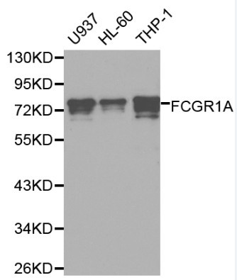
Anti-FCGR1A Antibody144-60077
ApplicationsWestern Blot, ImmunoHistoChemistry
ReactivityHuman, Mouse, Rat
TargetFCGR1A
- SizePrice
Product group Antibodies
ApplicationsFlow Cytometry, ImmunoHistoChemistry
ReactivityHuman
TargetFCGR1A
- SizePrice
Product group Antibodies

Anti-CD64 [H22]Ab00731-1.4
ApplicationsFlow Cytometry, ELISA, Other Application
ReactivityHuman
TargetFCGR1A
- SizePrice
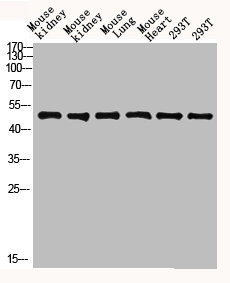
Product group Antibodies
Anti-FCGR1A AntibodyA29487
ApplicationsImmunoFluorescence, Western Blot, ImmunoHistoChemistry
ReactivityHuman
- SizePrice

Product group Antibodies
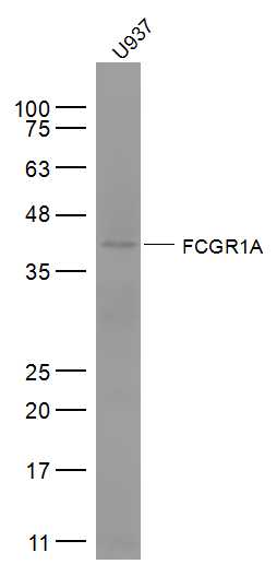
Anti-FCGR1A Antibody Picoband(r)A02428-1-CARRIER-FREE
ApplicationsFlow Cytometry, Western Blot, ELISA
ReactivityHuman, Rat
TargetFCGR1A
- SizePrice
Product group Antibodies

FCGR1A / CD64 Antibody (clone 10.1)LS-C770018
ApplicationsFlow Cytometry, ImmunoHistoChemistry
ReactivityHuman
TargetFCGR1A
- SizePrice
Product group Antibodies
FCGR1A AntibodyCSB-PA936888
ApplicationsWestern Blot, ELISA, ImmunoHistoChemistry
ReactivityHuman
TargetFCGR1A
- SizePrice
Product group Antibodies
FCGR1A/CD64 Polyclonal AntibodyBS-3511R
ApplicationsImmunoFluorescence, Western Blot, ELISA, ImmunoCytoChemistry, ImmunoHistoChemistry, ImmunoHistoChemistry Frozen, ImmunoHistoChemistry Paraffin
ReactivityHuman, Mouse, Rat
TargetFCGR1A
- SizePrice