


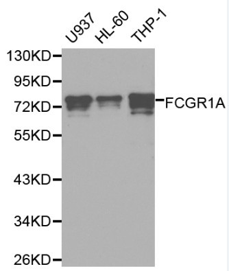
Figure 1. Western blot analysis of FCGR1A using anti-FCGR1A antibody (A02428-1). Electrophoresis was performed on a 5-20% SDS-PAGE gel at 70V (Stacking gel) / 90V (Resolving gel) for 2-3 hours. The sample well of each lane was loaded with 30 ug of sample under reducing conditions. Lane 1: human SiHa whole cell lysates, Lane 2: human A431 whole cell lysates, Lane 3: human HCCT tissue lysates, Lane 4: human Hela whole cell lysates, Lane 5: rat PC-12 whole cell lysates. After electrophoresis, proteins were transferred to a nitrocellulose membrane at 150 mA for 50-90 minutes. Blocked the membrane with 5% non-fat milk/TBS for 1.5 hour at RT. The membrane was incubated with rabbit anti-FCGR1A antigen affinity purified polyclonal antibody (Catalog # A02428-1) at 0.5 microg/mL overnight at 4°C, then washed with TBS-0.1%Tween 3 times with 5 minutes each and probed with a goat anti-rabbit IgG-HRP secondary antibody at a dilution of 1:5000 for 1.5 hour at RT. The signal is developed using an Enhanced Chemiluminescent detection (ECL) kit (Catalog # EK1002) with Tanon 5200 system. A specific band was detected for FCGR1A at approximately 70 kDa. The expected band size for FCGR1A is at 43 kDa.
. Overlay histogram showing U937 cells stained with A02428-1 (Blue line). To facilitate intracellular staining, cells were fixed with 4% paraformaldehyde and permeabilized with permeabilization buffer. The cells were blocked with 10% normal goat serum. And then incubated with rabbit anti-FCGR1A Antibody (A02428-1, 1 microg/1x106 cells) for 30 min at 20°C. DyLight®488 conjugated goat anti-rabbit IgG (BA1127, 5-10 microg/1x106 cells) was used as secondary antibody for 30 minutes at 20°C. Isotype control antibody (Green line) was rabbit IgG (1 microg/1x106) used under the same conditions. Unlabelled sample without incubation with primary antibody and secondary antibody (Red line) was used as a blank control.")
Figure 1. Western blot analysis of FCGR1A using anti-FCGR1A antibody (A02428-1). Electrophoresis was performed on a 5-20% SDS-PAGE gel at 70V (Stacking gel) / 90V (Resolving gel) for 2-3 hours. The sample well of each lane was loaded with 30 ug of sample under reducing conditions. Lane 1: human SiHa whole cell lysates, Lane 2: human A431 whole cell lysates, Lane 3: human HCCT tissue lysates, Lane 4: human Hela whole cell lysates, Lane 5: rat PC-12 whole cell lysates. After electrophoresis, proteins were transferred to a nitrocellulose membrane at 150 mA for 50-90 minutes. Blocked the membrane with 5% non-fat milk/TBS for 1.5 hour at RT. The membrane was incubated with rabbit anti-FCGR1A antigen affinity purified polyclonal antibody (Catalog # A02428-1) at 0.5 microg/mL overnight at 4°C, then washed with TBS-0.1%Tween 3 times with 5 minutes each and probed with a goat anti-rabbit IgG-HRP secondary antibody at a dilution of 1:5000 for 1.5 hour at RT. The signal is developed using an Enhanced Chemiluminescent detection (ECL) kit (Catalog # EK1002) with Tanon 5200 system. A specific band was detected for FCGR1A at approximately 70 kDa. The expected band size for FCGR1A is at 43 kDa.
Anti-FCGR1A Antibody Picoband(r)
A02428-1-CARRIER-FREE
ApplicationsFlow Cytometry, Western Blot, ELISA
Product group Antibodies
ReactivityHuman, Rat
TargetFCGR1A
Overview
- SupplierBoster Bio
- Product NameAnti-FCGR1A Antibody Picoband(r)
- Delivery Days Customer9
- ApplicationsFlow Cytometry, Western Blot, ELISA
- CertificationResearch Use Only
- ClonalityPolyclonal
- Concentration500 ug/ml
- Gene ID2209
- Target nameFCGR1A
- Target descriptionFc gamma receptor Ia
- Target synonymsCD64, CD64A, FCG1, FCGR1, FCRI, FcgammaRI, IGFR1, high affinity immunoglobulin gamma Fc receptor I, Fc fragment of IgG receptor Ia, Fc fragment of IgG, high affinity Ia, receptor (CD64), Fc fragment of IgG, high affinity Ia, receptor for (CD64), Fc-gamma RI, Fc-gamma receptor I A1, IgG Fc receptor I, fc-gamma RIA, fcgammaRIa
- HostRabbit
- IsotypeIgG
- Protein IDP12314
- Protein NameHigh affinity immunoglobulin gamma Fc receptor I
- Scientific DescriptionBoster Bio Anti-FCGR1A Antibody Picoband® catalog # A02428-1. Tested in ELISA, Flow Cytometry, WB applications. This antibody reacts with Human, Rat. The brand Picoband indicates this is a premium antibody that guarantees superior quality, high affinity, and strong signals with minimal background in Western blot applications. Only our best-performing antibodies are designated as Picoband, ensuring unmatched performance.
- ReactivityHuman, Rat
- Storage Instruction-20°C,2°C to 8°C
- UNSPSC12352203
Related products
Product group Antibodies
Anti-FCGR1A AntibodyA29487
ApplicationsImmunoFluorescence, Western Blot, ImmunoHistoChemistry
ReactivityHuman
- SizePrice
Product group Antibodies
Anti-FCGR1A Antibody144-60077
ApplicationsWestern Blot, ImmunoHistoChemistry
ReactivityHuman, Mouse, Rat
TargetFCGR1A
- SizePrice
Product group Antibodies
anti-CD64 (human), mAb (10.1) (Fab) (Biotin)ANC-216-530
ApplicationsFlow Cytometry, Western Blot, Neutralisation/Blocking
ReactivityHuman, Primate
TargetFCGR1A
- SizePrice
Product group Antibodies

FCGR1A / CD64 Antibody (clone 10.1)LS-C770018
ApplicationsFlow Cytometry, ImmunoHistoChemistry
ReactivityHuman
TargetFCGR1A
- SizePrice
Product group Antibodies

Anti-CD64 [H22]Ab00731-1.4
ApplicationsFlow Cytometry, ELISA, Other Application
ReactivityHuman
TargetFCGR1A
- SizePrice
Product group Antibodies
FCGR1A/CD64 Polyclonal AntibodyBS-3511R
ApplicationsImmunoFluorescence, Western Blot, ELISA, ImmunoCytoChemistry, ImmunoHistoChemistry, ImmunoHistoChemistry Frozen, ImmunoHistoChemistry Paraffin
ReactivityHuman, Mouse, Rat
TargetFCGR1A
- SizePrice
Product group Antibodies
ApplicationsWestern Blot, ELISA, ImmunoCytoChemistry, ImmunoHistoChemistry, ImmunoHistoChemistry Frozen, ImmunoHistoChemistry Paraffin
TargetFCGR1A
- SizePrice
Product group Antibodies
FCGR1A AntibodyCSB-PA936888
ApplicationsWestern Blot, ELISA, ImmunoHistoChemistry
ReactivityHuman
TargetFCGR1A
- SizePrice
Product group Antibodies
CD64 antibody [10.1] (PE-Cy7)GTX00467-10
ApplicationsFlow Cytometry
ReactivityHuman, Primate
TargetFCGR1A
- SizePrice