

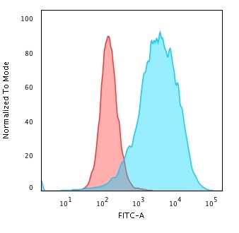
FACS analysis of PFA-fixed K562 cells using GTX02714 CD43 antibody [SPN/2049R]. Blue : Primary antibody Red : Isotype control
![ICC/IF analysis of K562 cells using GTX02714 CD43 antibody [SPN/2049R]. Green : Primary antibody Red : nuclear counterstain](https://www.genetex.com/upload/website/prouct_img/normal/GTX02714/GTX02714_20210319_ICCIF_w_23053122_687.webp "ICC/IF analysis of K562 cells using GTX02714 CD43 antibody [SPN/2049R]. Green : Primary antibody Red : nuclear counterstain")
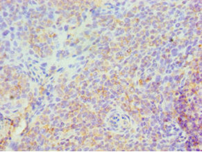
![IHC-P analysis of human tonsil tissue section using GTX02714 CD43 antibody [SPN/2049R].](https://www.genetex.com/upload/website/prouct_img/normal/GTX02714/GTX02714_20210319_IHC-P_w_23053122_626.webp "IHC-P analysis of human tonsil tissue section using GTX02714 CD43 antibody [SPN/2049R].")
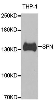
![WB analysis of K562 whole cell lysate using GTX02714 CD43 antibody [SPN/2049R].](https://www.genetex.com/upload/website/prouct_img/normal/GTX02714/GTX02714_20210319_WB_w_23053122_926.webp "WB analysis of K562 whole cell lysate using GTX02714 CD43 antibody [SPN/2049R].")
FACS analysis of PFA-fixed K562 cells using GTX02714 CD43 antibody [SPN/2049R]. Blue : Primary antibody Red : Isotype control
CD43 antibody [SPN/2049R]
GTX02714
ApplicationsFlow Cytometry, ImmunoFluorescence, Western Blot, ImmunoCytoChemistry, ImmunoHistoChemistry, ImmunoHistoChemistry Paraffin
Product group Antibodies
ReactivityHuman
TargetSPN
Overview
- SupplierGeneTex
- Product NameCD43 antibody [SPN/2049R]
- Delivery Days Customer9
- Application Supplier NoteWB: 1-2 microg/ml. ICC/IF: 1-2 microg/ml. IHC-P: 1-2 microg/ml. FACS: 1-2 microg/million cells. *Optimal dilutions/concentrations should be determined by the researcher.Not tested in other applications.
- ApplicationsFlow Cytometry, ImmunoFluorescence, Western Blot, ImmunoCytoChemistry, ImmunoHistoChemistry, ImmunoHistoChemistry Paraffin
- CertificationResearch Use Only
- ClonalityMonoclonal
- Clone IDSPN/2049R
- Concentration200 ug/ml
- ConjugateUnconjugated
- Gene ID6693
- Target nameSPN
- Target descriptionsialophorin
- Target synonymsCD43, GALGP, GPL115, LEU-22, LSN, leukosialin, galactoglycoprotein, leukocyte sialoglycoprotein, sialophorin (gpL115, leukosialin, CD43)
- HostRabbit
- IsotypeIgG
- Protein IDP16150
- Protein NameLeukosialin
- Scientific DescriptionThis gene encodes a highly sialylated glycoprotein that functions in antigen-specific activation of T cells, and is found on the surface of thymocytes, T lymphocytes, monocytes, granulocytes, and some B lymphocytes. It contains a mucin-like extracellular domain, a transmembrane region and a carboxy-terminal intracellular region. The extracellular domain has a high proportion of serine and threonine residues, allowing extensive O-glycosylation, and has one potential N-glycosylation site, while the carboxy-terminal region has potential phosphorylation sites that may mediate transduction of activation signals. Different glycoforms of this protein have been described. In stimulated immune cells, proteolytic cleavage of the extracellular domain occurs in some cell types, releasing a soluble extracellular fragment. Defects in expression of this gene are associated with Wiskott-Aldrich syndrome. [provided by RefSeq, Sep 2017]
- ReactivityHuman
- Storage Instruction2°C to 8°C
- UNSPSC12352203
Datasheet
Related products

Product group Antibodies

SPN / CD43 AntibodyABX027790
ApplicationsWestern Blot, ELISA, ImmunoHistoChemistry
- SizePrice
Product group Antibodies
Anti-SPN Antibody144-06412
ApplicationsWestern Blot, ImmunoHistoChemistry
ReactivityHuman, Mouse
TargetSPN
- SizePrice
Product group Antibodies
CD43 Recombinant AntibodyBSM-60237R
ApplicationsFlow Cytometry, Western Blot
ReactivityHuman
TargetSPN
- SizePrice
Product group Antibodies
SPN AntibodyCSB-PA022590ESR1HU
ApplicationsELISA, ImmunoHistoChemistry
ReactivityHuman
TargetSPN
- SizePrice
Product group Antibodies
Anti-SPN AntibodyA31363
ApplicationsWestern Blot, ImmunoHistoChemistry
ReactivityHuman
- SizePrice
Product group Antibodies

SPN / CD43 AntibodyLS-C835054
ApplicationsImmunoHistoChemistry
ReactivityHuman
TargetSPN
- SizePrice
![IHC-P analysis of human spleen tissue using GTX29088 CD43 antibody [MEM-59].](https://www.genetex.com/upload/website/prouct_img/normal/GTX29088/GTX29088_20191025_AP_003_223_w_23060722_215.webp)
Product group Antibodies
CD43 antibody [MEM-59]GTX29088
ApplicationsFlow Cytometry, ImmunoPrecipitation, Western Blot, ImmunoHistoChemistry, ImmunoHistoChemistry Paraffin
ReactivityHuman
TargetSPN
- SizePrice
![IHC-P analysis of human spleen tissue using GTX34507 CD43 antibody [SPM503].](https://www.genetex.com/upload/website/prouct_img/normal/GTX34507/GTX34507_20200115_IHC-P_1162_w_23060801_117.webp)
Product group Antibodies
CD43 antibody [SPM503]GTX34507
ApplicationsFlow Cytometry, ImmunoFluorescence, Western Blot, ImmunoCytoChemistry, ImmunoHistoChemistry, ImmunoHistoChemistry Paraffin
ReactivityHuman
TargetSPN
- SizePrice