

![CD43(DF-T1), Biotin conjugate, 0.1mg/mL [26628-22-8]](https://biotium.com/wp-content/uploads/2016/12/BNUB0027-1-1.jpg "CD43(DF-T1), Biotin conjugate, 0.1mg/mL [26628-22-8]")
CD43(DF-T1), Biotin conjugate, 0.1mg/mL [26628-22-8]
BNCB0027
ApplicationsFlow Cytometry, ImmunoFluorescence, Western Blot, ImmunoHistoChemistry, ImmunoHistoChemistry Paraffin
Product group Antibodies
ReactivityBovine, Human, Mouse
TargetSPN
Overview
- SupplierBiotium
- Product NameCD43(DF-T1), Biotin conjugate, 0.1mg/mL [26628-22-8]
- Delivery Days Customer9
- ApplicationsFlow Cytometry, ImmunoFluorescence, Western Blot, ImmunoHistoChemistry, ImmunoHistoChemistry Paraffin
- CAS Number26628-22-8
- CertificationResearch Use Only
- ClonalityMonoclonal
- Clone IDDF-T1
- Concentration0.1 mg/ml
- ConjugateBiotin
- Gene ID6693
- Target nameSPN
- Target descriptionsialophorin
- Target synonymsCD43, GALGP, GPL115, LEU-22, LSN, leukosialin, galactoglycoprotein, leukocyte sialoglycoprotein, sialophorin (gpL115, leukosialin, CD43)
- HostMouse
- IsotypeIgG1
- Protein IDP16150
- Protein NameLeukosialin
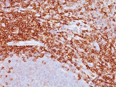
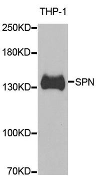
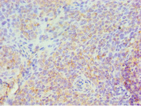
- Scientific DescriptionThis antibody recognizes a cell surface glycoprotein of 95/115/135 kDa (depending upon the extent of glycosylation), identified as CD43 . Epitope of MAb Bra7G is clearly different from that of MAb DF-T1, called b as opposed to a for DF-T1. 70-90% of T-cell lymphomas and from 22-37% of B-cell lymphomas express CD43. No reactivity has been observed with reactive B-cells. So a B-lineage population that co-expresses CD43 is highly likely to be a malignant lymphoma, especially a low-grade lymphoma, rather than a reactive B-cell population. When CD43 antibody is used in combination with anti-CD20, effective immunophenotyping of the lymphomas in formalin-fixed tissues can be obtained. Co-staining of a lymphoid infiltrate with anti-CD20 and anti-CD43 argues against a reactive process and favors a diagnosis of lymphoma.Primary antibodies are available purified, or with a selection of fluorescent CF® Dyes and other labels. CF® Dyes offer exceptional brightness and photostability. Note: Conjugates of blue fluorescent dyes like CF®405S and CF®405M are not recommended for detecting low abundance targets, because blue dyes have lower fluorescence and can give higher non-specific background than other dye colors.
- SourceAnimal
- ReactivityBovine, Human, Mouse
- Storage Instruction2°C to 8°C,RT
- UNSPSC41116161
MSDS
Related products
Product group Antibodies
Anti-SPN AntibodyA31363
ApplicationsWestern Blot, ImmunoHistoChemistry
ReactivityHuman
- SizePrice

Product group Antibodies
ApplicationsImmunoCytoChemistry
ReactivityHuman
TargetSPN
- SizePrice
Product group Antibodies
Anti-SPN Antibody144-06412
ApplicationsWestern Blot, ImmunoHistoChemistry
ReactivityHuman, Mouse
TargetSPN
- SizePrice
Product group Antibodies

SPN / CD43 AntibodyABX027790
ApplicationsWestern Blot, ELISA, ImmunoHistoChemistry
- SizePrice

Product group Antibodies
CD43 Recombinant AntibodyBSM-60237R
ApplicationsFlow Cytometry, Western Blot
ReactivityHuman
TargetSPN
- SizePrice
Product group Antibodies
SPN AntibodyCSB-PA022590ESR1HU
ApplicationsELISA, ImmunoHistoChemistry
ReactivityHuman
TargetSPN
- SizePrice
Product group Antibodies

Mouse anti MT1 / CD43MUB1203P
ApplicationsWestern Blot, ImmunoHistoChemistry, ImmunoHistoChemistry Frozen, ImmunoHistoChemistry Paraffin
ReactivityHuman
TargetSPN
- SizePrice
![FACS analysis of human peripheral blood using GTX78289 CD43 antibody [MEM-59] (FITC).](https://www.genetex.com/upload/website/prouct_img/normal/GTX78289/GTX78289_20191025_AP_006_274_w_23061322_748.webp)
Product group Antibodies
CD43 antibody [MEM-59] (FITC)GTX78289
ApplicationsFlow Cytometry
ReactivityHuman
TargetSPN
- SizePrice

Product group Antibodies
Anti-SPN AntibodyHPA055244
ApplicationsImmunoHistoChemistry
ReactivityHuman
TargetSPN
- SizePrice