


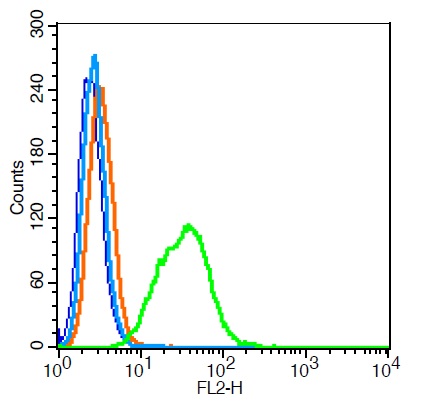
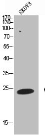
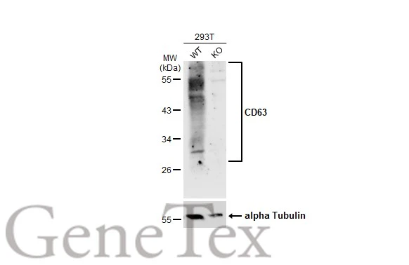
![CD63(MX-49.129.5), Biotin conjugate, 0.1mg/mL [26628-22-8]](https://biotium.com/wp-content/uploads/2016/12/BNUB0525-1-1.jpg "CD63(MX-49.129.5), Biotin conjugate, 0.1mg/mL [26628-22-8]")
![CD63(MX-49.129.5), Biotin conjugate, 0.1mg/mL [26628-22-8]](https://biotium.com/wp-content/uploads/2016/12/BNUB0525-2-1.jpg "CD63(MX-49.129.5), Biotin conjugate, 0.1mg/mL [26628-22-8]")
![CD63(MX-49.129.5), Biotin conjugate, 0.1mg/mL [26628-22-8]](https://biotium.com/wp-content/uploads/2016/12/BNUB0525-3-1.jpg "CD63(MX-49.129.5), Biotin conjugate, 0.1mg/mL [26628-22-8]")
![CD63(MX-49.129.5), Biotin conjugate, 0.1mg/mL [26628-22-8]](https://biotium.com/wp-content/uploads/2016/12/BNUB0525-4-1.jpg "CD63(MX-49.129.5), Biotin conjugate, 0.1mg/mL [26628-22-8]")
![CD63(MX-49.129.5), Biotin conjugate, 0.1mg/mL [26628-22-8]](https://biotium.com/wp-content/uploads/2016/12/BNUB0525-5-1.jpg "CD63(MX-49.129.5), Biotin conjugate, 0.1mg/mL [26628-22-8]")
![CD63(MX-49.129.5), Biotin conjugate, 0.1mg/mL [26628-22-8]](https://biotium.com/wp-content/uploads/2016/12/CD63-CF488A-MX-49.129.5-MCF7.jpg "CD63(MX-49.129.5), Biotin conjugate, 0.1mg/mL [26628-22-8]")
![CD63(MX-49.129.5), Biotin conjugate, 0.1mg/mL [26628-22-8]](https://biotium.com/wp-content/uploads/2016/12/CD63-CF568-MX-49.129.5-MCF7.jpg "CD63(MX-49.129.5), Biotin conjugate, 0.1mg/mL [26628-22-8]")
![CD63(MX-49.129.5), Biotin conjugate, 0.1mg/mL [26628-22-8]](https://biotium.com/wp-content/uploads/2016/12/CD63-CF568-MX-49.129.5-Jurkat.jpg "CD63(MX-49.129.5), Biotin conjugate, 0.1mg/mL [26628-22-8]")
![CD63(MX-49.129.5), Biotin conjugate, 0.1mg/mL [26628-22-8]](https://biotium.com/wp-content/uploads/2016/12/CD63-RPE-MX-49.129.5-Jurkat.jpg "CD63(MX-49.129.5), Biotin conjugate, 0.1mg/mL [26628-22-8]")
CD63(MX-49.129.5), Biotin conjugate, 0.1mg/mL [26628-22-8]
BNCB0525
ApplicationsFlow Cytometry, ImmunoFluorescence, ImmunoPrecipitation, Western Blot, ImmunoHistoChemistry, ImmunoHistoChemistry Paraffin
Product group Antibodies
ReactivityBovine, Human, Mouse
TargetCD63
Overview
- SupplierBiotium
- Product NameCD63(MX-49.129.5), Biotin conjugate, 0.1mg/mL [26628-22-8]
- Delivery Days Customer9
- ApplicationsFlow Cytometry, ImmunoFluorescence, ImmunoPrecipitation, Western Blot, ImmunoHistoChemistry, ImmunoHistoChemistry Paraffin
- CAS Number26628-22-8
- CertificationResearch Use Only
- ClonalityMonoclonal
- Clone IDMX-49.129.5
- Concentration0.1 mg/ml
- ConjugateBiotin
- Gene ID967
- Target nameCD63
- Target descriptionCD63 molecule
- Target synonymsAD1, HOP-26, ME491, MLA1, OMA81H, Pltgp40, TSPAN30, CD63 antigen, AD1 antigen, CD63 antigen (melanoma 1 antigen), granulophysin, limp1, melanoma-associated antigen ME491, melanoma-associated antigen MLA1, ocular melanoma-associated antigen, tetraspanin-30, tspan-30
- HostMouse
- IsotypeIgG1
- Protein IDP08962
- Protein NameCD63 antigen
- Scientific DescriptionThis MAb recognizes protein of 26 kDa-60 kDa, which is identified as CD63. Its epitope is different from that of MAb LAMP3/529. The tetraspanins are integral membrane proteins expressed on cell surface and granular membranes of hematopoietic cells and are components of multi-molecular complexes with specific integrins. The tetraspanin CD63 is a lysosomal membrane glycoprotein that translocates to the plasma membrane after platelet activation. CD63 is expressed on activated platelets, monocytes and macrophages, and is weakly expressed on granulocytes, T cell and B cells. It is located on the basophilic granule membranes and on the plasma membranes of lymphocytes and granulocytes. CD63 is a member of the TM4 superfamily of leukocyte glycoproteins that includes CD9, CD37 and CD53, which contain four transmembrane regions. CD63 may play a role in phagocytic and intracellular lysosome-phagosome fusion events. CD63 deficiency is associated with Hermansky-Pudlak syndrome and is strongly expressed during the early stages of melanoma progression. Primary antibodies are available purified, or with a selection of fluorescent CF® Dyes and other labels. CF® Dyes offer exceptional brightness and photostability. Note: Conjugates of blue fluorescent dyes like CF®405S and CF®405M are not recommended for detecting low abundance targets, because blue dyes have lower fluorescence and can give higher non-specific background than other dye colors.
- SourceAnimal
- ReactivityBovine, Human, Mouse
- Storage Instruction2°C to 8°C,RT
- UNSPSC41116161
MSDS
Related products
Product group Antibodies

Anti-CD63 [MOF11]Ab00388-1.1
ApplicationsFlow Cytometry
ReactivityHuman
TargetCD63
- SizePrice

Product group Antibodies
Anti-CD63 AntibodyA101276
ApplicationsELISA, ImmunoHistoChemistry
ReactivityHuman
- SizePrice
Product group Antibodies
References
CD63 Polyclonal AntibodyBS-1523R
ApplicationsFlow Cytometry, ImmunoFluorescence, Western Blot, ELISA, ImmunoCytoChemistry, ImmunoHistoChemistry, ImmunoHistoChemistry Frozen, ImmunoHistoChemistry Paraffin
ReactivityHuman
TargetCD63
- SizePrice
Product group Antibodies
CD63 AntibodyCSB-PA006039
ApplicationsWestern Blot, ELISA
ReactivityHuman
TargetCD63
- SizePrice
Product group Antibodies
ApplicationsFlow Cytometry
TargetCD63
- SizePrice
Product group Antibodies

ApplicationsImmunoFluorescence, ELISA, ImmunoHistoChemistry, ImmunoHistoChemistry Frozen, ImmunoHistoChemistry Paraffin
ReactivityHuman
TargetCD63
- SizePrice
Product group Antibodies
CD63 antibodyGTX132953
ApplicationsWestern Blot, ImmunoHistoChemistry, ImmunoHistoChemistry Paraffin
ReactivityHuman
TargetCD63
- SizePrice
Product group Antibodies

CD63 Antibody (Preservative Free)LS-C342960
ApplicationsWestern Blot, ELISA
ReactivityHuman
TargetCD63
- SizePrice