

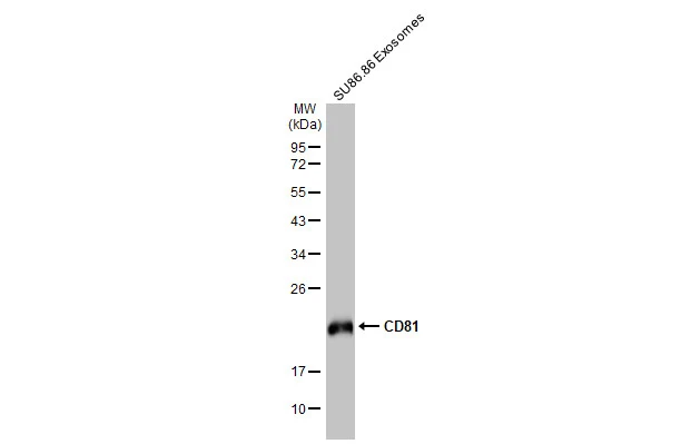
SU86.86 exsosome extracts (1 μg) was separated by 12% SDS-PAGE, and the membrane was blotted with CD81 antibody [HL1666] (GTX637264) diluted at 1:5000. The HRP-conjugated anti-rabbit IgG antibody (GTX213110-01) was used to detect the primary antibody.
![Jurkat membrane extract (30 μg) was separated by 12% SDS-PAGE, and the membrane was blotted with CD81 antibody [HL1666] (GTX637264) diluted at 1:500. The HRP-conjugated anti-rabbit IgG antibody (GTX213110-01) was used to detect the primary antibody.](https://www.genetex.com/upload/website/prouct_img/normal/GTX637264/GTX637264_44823_20221104_WB_Fraction_22110919_753.webp "Jurkat membrane extract (30 μg) was separated by 12% SDS-PAGE, and the membrane was blotted with CD81 antibody [HL1666] (GTX637264) diluted at 1:500. The HRP-conjugated anti-rabbit IgG antibody (GTX213110-01) was used to detect the primary antibody.")
![CD81 antibody [HL1666] detects CD81 protein by immunofluorescent analysis. Sample: Jurkat cells were fixed in 4% paraformaldehyde at RT for 15 min. Green: CD81 stained by CD81 antibody [HL1666] (GTX637264) diluted at 1:500. Blue: Fluoroshield with DAPI (GTX30920).](https://www.genetex.com/upload/website/prouct_img/normal/GTX637264/GTX637264_44823_20221230_ICC_IF_23010219_878.webp "CD81 antibody [HL1666] detects CD81 protein by immunofluorescent analysis. Sample: Jurkat cells were fixed in 4% paraformaldehyde at RT for 15 min. Green: CD81 stained by CD81 antibody [HL1666] (GTX637264) diluted at 1:500. Blue: Fluoroshield with DAPI (GTX30920).")
![Whole cell extract (30 μg) was separated by 12% SDS-PAGE, and the membrane was blotted with CD81 antibody [HL1666] (GTX637264) diluted at 1:1000. The HRP-conjugated anti-rabbit IgG antibody (GTX213110-01) was used to detect the primary antibody.](https://www.genetex.com/upload/website/prouct_img/normal/GTX637264/GTX637264_44823_20231117_WB_M_23112021_524.webp "Whole cell extract (30 μg) was separated by 12% SDS-PAGE, and the membrane was blotted with CD81 antibody [HL1666] (GTX637264) diluted at 1:1000. The HRP-conjugated anti-rabbit IgG antibody (GTX213110-01) was used to detect the primary antibody.")
![Whole cell extract (30 μg) was separated by 12% SDS-PAGE, and the membrane was blotted with CD81 antibody [HL1666] (GTX637264) diluted at 1:1000. The HRP-conjugated anti-rabbit IgG antibody (GTX213110-01) was used to detect the primary antibody.](https://www.genetex.com/upload/website/prouct_img/normal/GTX637264/GTX637264_44823_20231117_WB_R_23112021_525.webp "Whole cell extract (30 μg) was separated by 12% SDS-PAGE, and the membrane was blotted with CD81 antibody [HL1666] (GTX637264) diluted at 1:1000. The HRP-conjugated anti-rabbit IgG antibody (GTX213110-01) was used to detect the primary antibody.")
![Non-transfected (–) and transfected (+) 293T whole cell extracts (30 μg) were separated by 12% SDS-PAGE, and the membrane was blotted with CD81 antibody [HL1666] (GTX637264) diluted at 1:10000. The HRP-conjugated anti-rabbit IgG antibody (GTX213110-01) was used to detect the primary antibody.](https://www.genetex.com/upload/website/prouct_img/normal/GTX637264/GTX637264_45488_20240802_WB_B_24110700_452.webp "Non-transfected (–) and transfected (+) 293T whole cell extracts (30 μg) were separated by 12% SDS-PAGE, and the membrane was blotted with CD81 antibody [HL1666] (GTX637264) diluted at 1:10000. The HRP-conjugated anti-rabbit IgG antibody (GTX213110-01) was used to detect the primary antibody.")
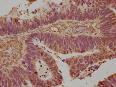
![CD81 antibody [HL1666] detects CD81 protein by immunohistochemical analysis. Sample: Paraffin-embedded mouse tissues. CD81 stained by CD81 antibody [HL1666] (GTX637264) diluted at 1:3200. Antigen Retrieval: Citrate buffer, pH 6.0, 15 min](https://www.genetex.com/upload/website/prouct_img/normal/GTX637264/GTX637264_45488_20250731_IHC-P_M_Multiple_RPKM_25081423_973.webp "CD81 antibody [HL1666] detects CD81 protein by immunohistochemical analysis. Sample: Paraffin-embedded mouse tissues. CD81 stained by CD81 antibody [HL1666] (GTX637264) diluted at 1:3200. Antigen Retrieval: Citrate buffer, pH 6.0, 15 min")
![CD81 antibody [HL1666] detects CD81 protein by immunohistochemical analysis. Sample: Paraffin-embedded mouse heart. CD81 stained by CD81 antibody [HL1666] (GTX637264) diluted at 1:3200. Antigen Retrieval: Citrate buffer, pH 6.0, 15 min](https://www.genetex.com/upload/website/prouct_img/normal/GTX637264/GTX637264_45488_20250731_IHC-P_M_25081423_601.webp "CD81 antibody [HL1666] detects CD81 protein by immunohistochemical analysis. Sample: Paraffin-embedded mouse heart. CD81 stained by CD81 antibody [HL1666] (GTX637264) diluted at 1:3200. Antigen Retrieval: Citrate buffer, pH 6.0, 15 min")
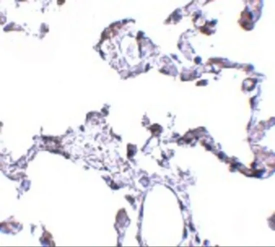
![CD81 antibody [HL1666] detects CD81 protein by immunohistochemical analysis. Sample: Paraffin-embedded mouse lung. CD81 stained by CD81 antibody [HL1666] (GTX637264) diluted at 1:3200. Antigen Retrieval: Citrate buffer, pH 6.0, 15 min](https://www.genetex.com/upload/website/prouct_img/normal/GTX637264/GTX637264_45488_20250731_IHC-P_M_2_25081423_274.webp "CD81 antibody [HL1666] detects CD81 protein by immunohistochemical analysis. Sample: Paraffin-embedded mouse lung. CD81 stained by CD81 antibody [HL1666] (GTX637264) diluted at 1:3200. Antigen Retrieval: Citrate buffer, pH 6.0, 15 min")
![CD81 antibody [HL1666] detects CD81 protein by immunohistochemical analysis. Sample: Paraffin-embedded rat salivary gland. CD81 stained by CD81 antibody [HL1666] (GTX637264) diluted at 1:3000. Antigen Retrieval: Citrate buffer, pH 6.0, 15 min](https://www.genetex.com/upload/website/prouct_img/normal/GTX637264/GTX637264_45488_20250925_IHC-P_R_25100122_547.webp "CD81 antibody [HL1666] detects CD81 protein by immunohistochemical analysis. Sample: Paraffin-embedded rat salivary gland. CD81 stained by CD81 antibody [HL1666] (GTX637264) diluted at 1:3000. Antigen Retrieval: Citrate buffer, pH 6.0, 15 min")
SU86.86 exsosome extracts (1 μg) was separated by 12% SDS-PAGE, and the membrane was blotted with CD81 antibody [HL1666] (GTX637264) diluted at 1:5000. The HRP-conjugated anti-rabbit IgG antibody (GTX213110-01) was used to detect the primary antibody.
CD81 antibody [HL1666]
GTX637264
ApplicationsImmunoFluorescence, Western Blot, ImmunoCytoChemistry, ImmunoHistoChemistry, ImmunoHistoChemistry Paraffin
Product group Antibodies
ReactivityHuman, Mouse, Rat
TargetCD81
Overview
- SupplierGeneTex
- Product NameCD81 antibody [HL1666]
- Delivery Days Customer9
- Application Supplier NoteWB: 1:1000-1:10000. *Optimal dilutions/concentrations should be determined by the researcher.Not tested in other applications.
- ApplicationsImmunoFluorescence, Western Blot, ImmunoCytoChemistry, ImmunoHistoChemistry, ImmunoHistoChemistry Paraffin
- CertificationResearch Use Only
- ClonalityMonoclonal
- Clone IDHL1666
- Concentration2 mg/ml
- ConjugateUnconjugated
- Gene ID975
- Target nameCD81
- Target descriptionCD81 molecule
- Target synonymsCVID6, S5.7, TAPA1, TSPAN28, CD81 antigen, 26 kDa cell surface protein TAPA-1, CD81 antigen (target of antiproliferative antibody 1), tetraspanin-28, tspan-28
- HostRabbit
- IsotypeIgG
- Protein IDP60033
- Protein NameCD81 antigen
- Scientific DescriptionThe protein encoded by this gene is a member of the transmembrane 4 superfamily, also known as the tetraspanin family. Most of these members are cell-surface proteins that are characterized by the presence of four hydrophobic domains. The proteins mediate signal transduction events that play a role in the regulation of cell development, activation, growth and motility. This encoded protein is a cell surface glycoprotein that is known to complex with integrins. This protein appears to promote muscle cell fusion and support myotube maintenance. Also it may be involved in signal transduction. This gene is localized in the tumor-suppressor gene region and thus it is a candidate gene for malignancies. Two transcript variants encoding different isoforms have been found for this gene. [provided by RefSeq, Jul 2014]
- ReactivityHuman, Mouse, Rat
- Storage Instruction-20°C or -80°C,2°C to 8°C
- UNSPSC41116161
Datasheet
Related products
Product group Antibodies
CD81 AntibodyCSB-PA004960NA01HU
ApplicationsELISA, ImmunoHistoChemistry
ReactivityHuman
TargetCD81
- SizePrice

Product group Antibodies
Anti-TAPA1/CD81 Antibody Picoband(r)A01281-2-CARRIER-FREE
ApplicationsFlow Cytometry, Western Blot, ELISA, ImmunoCytoChemistry, ImmunoHistoChemistry, ImmunoHistoChemistry Frozen
ReactivityHuman, Mouse, Rat
TargetCD81
- SizePrice
Product group Antibodies

Anti-CD81 [1D6]Ab00697-1.1
ApplicationsFunctional Assay, Flow Cytometry, ImmunoFluorescence, ImmunoPrecipitation, Western Blot, ELISA, ImmunoCytoChemistry, ImmunoHistoChemistry, ImmunoHistoChemistry Paraffin
ReactivityGoat, Human, Primate, Sheep
TargetCD81
- SizePrice

Product group Antibodies
Anti-CD81 AntibodyA98238
ApplicationsWestern Blot, ELISA
ReactivityHuman, Mouse, Rat
- SizePrice

Product group Antibodies
Anti-CD81 AntibodyHPA007234
ApplicationsImmunoCytoChemistry, ImmunoHistoChemistry
ReactivityHuman
TargetCD81
- SizePrice
Product group Antibodies

CD81 Antibody (PE)LS-C485188
ApplicationsFlow Cytometry
ReactivityHuman
TargetCD81
- SizePrice
Product group Antibodies
References
CD81 antibodyGTX31381
ApplicationsImmunoFluorescence, Western Blot, ELISA, ImmunoCytoChemistry, ImmunoHistoChemistry, ImmunoHistoChemistry Paraffin
ReactivityHuman, Mouse, Rat
TargetCD81
- SizePrice
Product group Antibodies
ApplicationsFlow Cytometry
TargetCD81
- SizePrice
Product group Antibodies
CD81 Polyclonal AntibodyBS-6934R
ApplicationsImmunoFluorescence, Western Blot, ELISA, ImmunoCytoChemistry, ImmunoHistoChemistry, ImmunoHistoChemistry Frozen, ImmunoHistoChemistry Paraffin
ReactivityHuman, Mouse, Rat
TargetCD81
- SizePrice