

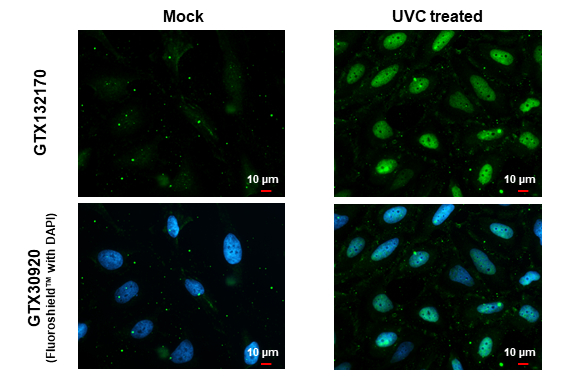
Chk1 (phospho Ser317) antibody detects Chk1 (phospho Ser317) protein at nucleus by immunofluorescent analysis. Sample: Mock and treated HeLa cells were fixed in 4% paraformaldehyde at RT for 15 min. Green: Chk1 (phospho Ser317) stained by Chk1 (phospho Ser317) antibody (GTX132170) diluted at 1:50. Red: alpha Tubulin, a cytoskeleton marker, stained by alpha Tubulin antibody [GT114] (GTX628802) diluted at 1:1000. Blue: Fluoroshield with DAPI (GTX30920).
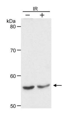
 and treated (+) HCT116 whole cell extracts (30 μg) were separated by 10% SDS-PAGE, and the membrane was blotted with Chk1 (phospho Ser317) antibody (GTX132170) diluted at 1:500.")
Chk1 (phospho Ser317) antibody detects Chk1 (phospho Ser317) protein at nucleus by immunofluorescent analysis. Sample: Mock and treated HeLa cells were fixed in 4% paraformaldehyde at RT for 15 min. Green: Chk1 (phospho Ser317) stained by Chk1 (phospho Ser317) antibody (GTX132170) diluted at 1:50. Red: alpha Tubulin, a cytoskeleton marker, stained by alpha Tubulin antibody [GT114] (GTX628802) diluted at 1:1000. Blue: Fluoroshield with DAPI (GTX30920).
Chk1 (phospho Ser317) antibody
GTX132170
ApplicationsImmunoFluorescence, Western Blot, ImmunoCytoChemistry
Product group Antibodies
ReactivityHuman
TargetCHEK1
Overview
- SupplierGeneTex
- Product NameChk1 (phospho Ser317) antibody
- Delivery Days Customer9
- Application Supplier NoteWB: 1:500-1:3000. ICC/IF: 1:100-1:1000. *Optimal dilutions/concentrations should be determined by the researcher.Not tested in other applications.
- ApplicationsImmunoFluorescence, Western Blot, ImmunoCytoChemistry
- CertificationResearch Use Only
- ClonalityPolyclonal
- Concentration2.2 mg/ml
- ConjugateUnconjugated
- Gene ID1111
- Target nameCHEK1
- Target descriptioncheckpoint kinase 1
- Target synonymsCHK1, OZEMA21, serine/threonine-protein kinase Chk1, CHK1 checkpoint homolog, Checkpoint, S. pombe, homolog of, 1, Chk1-S, cell cycle checkpoint kinase
- HostRabbit
- IsotypeIgG
- Protein IDO14757
- Protein NameSerine/threonine-protein kinase Chk1
- Scientific DescriptionRequired for checkpoint mediated cell cycle arrest in response to DNA damage or the presence of unreplicated DNA. May also negatively regulate cell cycle progression during unperturbed cell cycles. Recognizes the substrate consensus sequence [R-X-X-S/T]. Binds to and phosphorylates CDC25A, CDC25B and CDC25C. Phosphorylation of CDC25A at Ser-178 and Thr-507 and phosphorylation of CDC25C at Ser-216 creates binding sites for 14-3-3 proteins which inhibit CDC25A and CDC25C. Phosphorylation of CDC25A at Ser-76, Ser-124, Ser-178, Ser-279 and Ser-293 promotes proteolysis of CDC25A. Inhibition of CDC25 activity leads to increased inhibitory tyrosine phosphorylation of CDK-cyclin complexes and blocks cell cycle progression. Binds to and phosphorylates RAD51 at Thr-309, which may enhance the association of RAD51 with chromatin and promote DNA repair by homologous recombination. Binds to and phosphorylates TLK1 at Ser-743, which prevents the TLK1-dependent phosphorylation of the chromatin assembly factor ASF1A. This may affect chromatin assembly during S phase or DNA repair. May also phosphorylate multiple sites within the C-terminus of TP53, which promotes activation of TP53 by acetylation and enhances suppression of cellular proliferation.
- ReactivityHuman
- Storage Instruction-20°C or -80°C,2°C to 8°C
- UNSPSC41116161
Datasheet
Related products
Product group Antibodies
Anti-Chk1 AntibodyA100008
ApplicationsImmunoFluorescence, Western Blot, ELISA
ReactivityHuman
- SizePrice

Product group Antibodies
Anti-Chk1/CHEK1 Antibody Picoband(r)A01060-CARRIER-FREE
ApplicationsFlow Cytometry, Western Blot
ReactivityHuman
TargetCHEK1
- SizePrice
Product group Antibodies
Anti-CHEK1 Antibody144-07653
ApplicationsImmunoFluorescence, Western Blot, ImmunoHistoChemistry
ReactivityHuman, Mouse, Rat
TargetCHEK1
- SizePrice
Product group Antibodies

CHEK1 / CHK1 AntibodyLS-C761161
ApplicationsWestern Blot, ImmunoHistoChemistry
ReactivityBovine, Human, Mouse, Rat
TargetCHEK1
- SizePrice
Product group Antibodies
References
ApplicationsImmunoFluorescence, ELISA, ImmunoCytoChemistry, ImmunoHistoChemistry, ImmunoHistoChemistry Frozen, ImmunoHistoChemistry Paraffin
ReactivityEquine, Human, Mouse, Porcine, Rabbit, Rat
TargetCHEK1
- SizePrice
Product group Antibodies
CHEK1 AntibodyCSB-PA001623
ApplicationsImmunoFluorescence, Western Blot, ELISA, ImmunoHistoChemistry
ReactivityHuman
TargetCHEK1
- SizePrice
Product group Antibodies

Goat anti-CHEK1EB08452
ApplicationsWestern Blot, ELISA
ReactivityBovine, Human, Mouse, Rat
TargetCHEK1
- SizePrice
Product group Antibodies
Chek1 Recombinant AntibodyCAC12088
ApplicationsImmunoFluorescence, Western Blot, ELISA, ImmunoHistoChemistry
TargetCHEK1
- SizePrice
Product group Antibodies
Chk1 antibodyGTX22845
ApplicationsWestern Blot, Other Application
ReactivityHuman
TargetCHEK1
- SizePrice
![Untreated (–) and treated (+) HCT116 whole cell extracts (30 μg) were separated by 10% SDS-PAGE, and the membrane was blotted with Chk1 antibody [ST57-09] (GTX01101) diluted at 1:500. The HRP-conjugated anti-rabbit IgG antibody (GTX213110-01) was used to detect the primary antibody.](https://www.genetex.com/upload/website/prouct_img/normal/GTX01101/GTX01101_HM0401_20220211_WB_treatment_Cisplatin_peptideblocking_w_23053121_692.webp)
Product group Antibodies
Chk1 antibody [ST57-09]GTX01101
ApplicationsWestern Blot
ReactivityHuman
TargetCHEK1
- SizePrice