


![Chromogranin A(CGA/414), CF405S conjugate, 0.1mg/mL [26628-22-8]](https://biotium.com/wp-content/uploads/2016/12/BNUB0414-1-1.jpg "Chromogranin A(CGA/414), CF405S conjugate, 0.1mg/mL [26628-22-8]")
Chromogranin A(CGA/414), CF405S conjugate, 0.1mg/mL [26628-22-8]
BNC040414
ApplicationsImmunoHistoChemistry, ImmunoHistoChemistry Paraffin
Product group Antibodies
TargetCHGA
Overview
- SupplierBiotium
- Product NameChromogranin A(CGA/414), CF405S conjugate, 0.1mg/mL
- Delivery Days Customer9
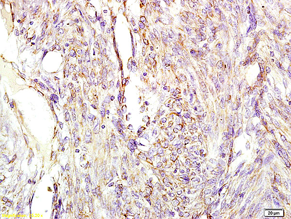
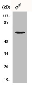
- ApplicationsImmunoHistoChemistry, ImmunoHistoChemistry Paraffin
- CertificationResearch Use Only
- ClonalityMonoclonal
- Clone IDCGA/414
- Concentration0.1 mg/ml
- ConjugateOther Conjugate
- Gene ID1113
- Target nameCHGA
- Target descriptionchromogranin A
- Target synonymsbetagranin (N-terminal fragment of chromogranin A); catestatin; CGA; chromofungin; chromogranin-A; parathyroid secretory protein 1; pituitary secretory protein I; SP-I
- HostMouse
- IsotypeIgG1
- Protein IDP10645
- Protein NameChromogranin-A
- Scientific DescriptionChromogranin A is present in neuroendocrine cells throughout the body, including the neuroendocrine cells of the large and small intestine, adrenal medulla and pancreatic islets. It is an excellent marker for carcinoid tumors, pheochromocytomas, paragangliomas, and other neuroendocrine tumors. Co-expression of chromogranin A and neuron specific enolase (NSE) is common in neuroendocrine neoplasms. Reportedly, co-expression of certain keratins and chromogranin indicates neuroendocrine lineage. The presence of strong anti-chromogranin staining and absence of anti-keratin staining should raise the possibility of paraganglioma. The co-expression of chromogranin and NSE is typical of neuroendocrine neoplasms. Most pituitary adenomas and prolactinomas readily express chromogranin.Primary antibodies are available purified, or with a selection of fluorescent CF® Dyes and other labels. CF® Dyes offer exceptional brightness and photostability. Note: Conjugates of blue fluorescent dyes like CF®405S and CF®405M are not recommended for detecting low abundance targets, because blue dyes have lower fluorescence and can give higher non-specific background than other dye colors.
- SourceAnimal
- Storage Instruction2°C to 8°C
- UNSPSC12352203
Related products
Product group Antibodies

ApplicationsFlow Cytometry, Western Blot, ELISA
TargetCHGA
- SizePrice
Product group Antibodies
Chga Polyclonal AntibodyCAC08310
ApplicationsELISA, ImmunoHistoChemistry
TargetCHGA
- SizePrice
Product group Antibodies
References
CHGA Polyclonal AntibodyBS-0539R
ApplicationsImmunoFluorescence, ELISA, ImmunoCytoChemistry, ImmunoHistoChemistry, ImmunoHistoChemistry Frozen, ImmunoHistoChemistry Paraffin
TargetCHGA
- SizePrice
Product group Antibodies
References
Chromogranin A antibodyGTX113165
ApplicationsWestern Blot, ImmunoHistoChemistry, ImmunoHistoChemistry Paraffin
TargetCHGA
- SizePrice
Product group Antibodies
CHGA AntibodyCSB-PA001643
ApplicationsWestern Blot, ELISA, ImmunoHistoChemistry
TargetCHGA
- SizePrice

Product group Antibodies
Anti-Chromogranin A/CHGA Antibody Picoband(r)A01256-4-CARRIER-FREE
ApplicationsFlow Cytometry, Western Blot, ELISA, ImmunoHistoChemistry
TargetCHGA
- SizePrice
Product group Antibodies
ApplicationsWestern Blot, ImmunoHistoChemistry
- SizePrice