

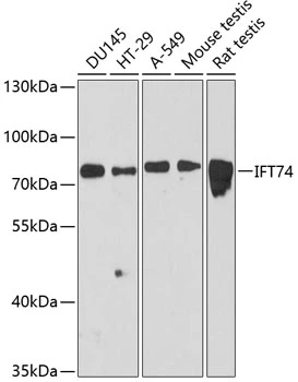
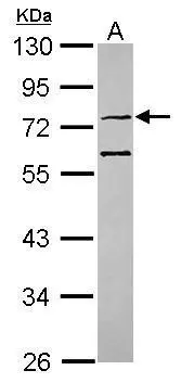
WB analysis of various sample lysates using GTX64834 CMG1 antibody. Dilution : 1:3000 Loading : 25μg per lane
WB analysis of various sample lysates using GTX64834 CMG1 antibody. Dilution : 1:3000 Loading : 25μg per lane
CMG1 antibody
GTX64834
ApplicationsWestern Blot
Product group Antibodies
ReactivityHuman, Mouse, Rat
TargetIFT74
Overview
- SupplierGeneTex
- Product NameCMG1 antibody
- Delivery Days Customer9
- Application Supplier NoteWB: 1:1000 - 1:3000. *Optimal dilutions/concentrations should be determined by the researcher.Not tested in other applications.
- ApplicationsWestern Blot
- CertificationResearch Use Only
- ClonalityPolyclonal
- ConjugateUnconjugated
- Gene ID80173
- Target nameIFT74
- Target descriptionintraflagellar transport 74
- Target synonymsBBS22, CCDC2, CMG-1, CMG1, JBTS40, SPGF58, intraflagellar transport protein 74 homolog, capillary morphogenesis gene 1 protein, capillary morphogenesis protein 1, coiled-coil domain containing 2, coiled-coil domain-containing protein 2, intraflagellar transport 74 homolog
- HostRabbit
- IsotypeIgG
- Protein IDQ96LB3
- Protein NameIntraflagellar transport protein 74 homolog
- Scientific DescriptionThis gene encodes a core intraflagellar transport (IFT) protein which belongs to a multi-protein complex involved in the transport of ciliary proteins along axonemal microtubules. IFT proteins are found at the base of the cilium as well as inside the cilium, where they assemble into long arrays between the ciliary base and tip. This protein, together with intraflagellar transport protein 81, binds and transports tubulin within cilia and is required for ciliogenesis. Naturally occurring mutations in this gene are associated with amyotrophic lateral sclerosis--frontotemporal dementia and Bardet-Biedl Syndrome. [provided by RefSeq, Mar 2017]
- ReactivityHuman, Mouse, Rat
- Storage Instruction-20°C or -80°C,2°C to 8°C
- UNSPSC41116161
Datasheet
Related products

Product group Antibodies
Anti-IFT74 Antibody Picoband(r)A08931-2-CARRIER-FREE
ApplicationsFlow Cytometry, Western Blot, ELISA, ImmunoHistoChemistry
ReactivityHuman, Monkey, Mouse, Rat
TargetIFT74
- SizePrice
Product group Antibodies
Anti-IFT74 Antibody144-12672
ApplicationsWestern Blot
ReactivityHuman, Mouse, Rat
TargetIFT74
- SizePrice
Product group Antibodies
ApplicationsWestern Blot, ELISA
ReactivityHuman
- SizePrice
Product group Antibodies

ApplicationsWestern Blot, ELISA
ReactivityHuman, Rat
TargetIFT74
- SizePrice

Product group Antibodies
Anti-IFT74 AntibodyHPA020247
ApplicationsImmunoHistoChemistry
ReactivityHuman
TargetIFT74
- SizePrice
Product group Antibodies
IFT74 AntibodyCSB-PA040376
ApplicationsELISA, ImmunoHistoChemistry
ReactivityHuman
TargetIFT74
- SizePrice
Product group Antibodies

IFT74 / CCDC2 AntibodyLS-C403075
ApplicationsWestern Blot, ELISA
ReactivityHuman
TargetIFT74
- SizePrice

Product group Antibodies
CMG1 Polyclonal AntibodyBS-7050R
ApplicationsImmunoFluorescence, ELISA, ImmunoCytoChemistry, ImmunoHistoChemistry, ImmunoHistoChemistry Frozen, ImmunoHistoChemistry Paraffin
ReactivityBovine, Equine, Human, Mouse, Porcine, Rabbit, Rat
TargetIFT74
- SizePrice
Product group Antibodies
CMG1 antibody [C3], C-termGTX106317
ApplicationsImmunoFluorescence, Western Blot, ImmunoCytoChemistry
ReactivityHuman
TargetIFT74
- SizePrice
Product group Antibodies
CMG1 antibody [N1N3]GTX118199
ApplicationsWestern Blot
ReactivityHuman
TargetIFT74
- SizePrice