

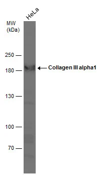
Whole cell extract (30 μg) was separated by 5% SDS-PAGE, and the membrane was blotted with Collagen III alpha1 antibody [C2C3-2], C-term (GTX111643) diluted at 1:1000.
![Collagen III antibody [C2C3-2], C-term detects Collagen III protein at cell membrane by immunofluorescent analysis. Sample: HeLa cells were fixed in ice-cold MeOH for 5 min. Green: Collagen III stained by Collagen III antibody [C2C3-2], C-term (GTX111643) diluted at 1:500.](https://www.genetex.com/upload/website/prouct_img/normal/GTX111643/GTX111643_44370_20210903_ICC_IF_w_23060500_347.webp "Collagen III antibody [C2C3-2], C-term detects Collagen III protein at cell membrane by immunofluorescent analysis. Sample: HeLa cells were fixed in ice-cold MeOH for 5 min. Green: Collagen III stained by Collagen III antibody [C2C3-2], C-term (GTX111643) diluted at 1:500.")
 antibody at 1:500 dilution.
Antigen Retrieval: Trilogy? (EDTA based, pH 8.0) buffer, 15min")

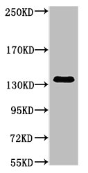
![Various whole cell extracts (30 μg) were separated by 5% SDS-PAGE, and the membrane was blotted with Collagen III antibody [C2C3-2], C-term (GTX111643) diluted at 1:2000. The HRP-conjugated anti-rabbit IgG antibody (GTX213110-01) was used to detect the primary antibody. Corresponding RNA expression data for the same cell lines are based on Human Protein Atlas program.](https://www.genetex.com/upload/website/prouct_img/normal/GTX111643/GTX111643_45331_20240301_WB_TPM_watermark_25061003_526.webp "Various whole cell extracts (30 μg) were separated by 5% SDS-PAGE, and the membrane was blotted with Collagen III antibody [C2C3-2], C-term (GTX111643) diluted at 1:2000. The HRP-conjugated anti-rabbit IgG antibody (GTX213110-01) was used to detect the primary antibody. Corresponding RNA expression data for the same cell lines are based on Human Protein Atlas program.")
Whole cell extract (30 μg) was separated by 5% SDS-PAGE, and the membrane was blotted with Collagen III alpha1 antibody [C2C3-2], C-term (GTX111643) diluted at 1:1000.
Collagen III antibody [C2C3-2], C-term
GTX111643
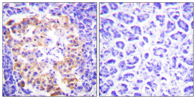
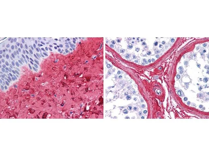
ApplicationsImmunoFluorescence, Western Blot, ImmunoCytoChemistry, ImmunoHistoChemistry, ImmunoHistoChemistry Paraffin
Product group Antibodies
ReactivityHuman
TargetCOL3A1
Overview
- SupplierGeneTex
- Product NameCollagen III antibody [C2C3-2], C-term
- Delivery Days Customer9
- Application Supplier NoteWB: 1:500-1:3000. ICC/IF: 1:100-1:1000. IHC-P: 1:100-1:1000. *Optimal dilutions/concentrations should be determined by the researcher.Not tested in other applications.
- ApplicationsImmunoFluorescence, Western Blot, ImmunoCytoChemistry, ImmunoHistoChemistry, ImmunoHistoChemistry Paraffin
- CertificationResearch Use Only
- ClonalityPolyclonal
- Concentration1.98 mg/ml
- ConjugateUnconjugated
- Gene ID1281
- Target nameCOL3A1
- Target descriptioncollagen type III alpha 1 chain
- Target synonymsEDS4A, EDSVASC, PMGEDSV, collagen alpha-1(III) chain, Ehlers-Danlos syndrome type IV, autosomal dominant, alpha-1 type III collagen, alpha1 (III) collagen, collagen, fetal, collagen, type III, alpha 1
- HostRabbit
- IsotypeIgG
- Protein IDP02461
- Protein NameCollagen alpha-1(III) chain
- Scientific DescriptionThis gene encodes the pro-alpha1 chains of type III collagen, a fibrillar collagen that is found in extensible connective tissues such as skin, lung, uterus, intestine and the vascular system, frequently in association with type I collagen. Mutations in this gene are associated with Ehlers-Danlos syndrome types IV, and with aortic and arterial aneurysms. Two transcripts, resulting from the use of alternate polyadenylation signals, have been identified for this gene. [provided by R. Dalgleish]
- ReactivityHuman
- Storage Instruction-20°C or -80°C,2°C to 8°C
- UNSPSC12352203
References
- Zheng Z, Ao X, Li P, et al. CRLF1 Is a Key Regulator in the Ligamentum Flavum Hypertrophy. Front Cell Dev Biol. 2020,8:858. doi: 10.3389/fcell.2020.00858Read this paper
- Yeh LC, Chen SP, Liao FH, et al. The Bioactive Core and Corona Synergism of Quantized Gold Enables Slowed Inflammation and Increased Tissue Regeneration in Wound Hypoxia. Int J Mol Sci. 2020,21(5). doi: 10.3390/ijms21051699Read this paper
- Shentu TP, Huang TS, Cernelc-Kohan M, et al. Thy-1 dependent uptake of mesenchymal stem cell-derived extracellular vesicles blocks myofibroblastic differentiation. Sci Rep. 2017,7(1):18052. doi: 10.1038/s41598-017-18288-9Read this paper
- Saito Y, Chikenji T, Ozasa Y, et al. PDGFR Signaling Mediates Hyperproliferation and Fibrotic Responses of Subsynovial Connective Tissue Cells in Idiopathic Carpal Tunnel Syndrome. Sci Rep. 2017,7(1):16192. doi: 10.1038/s41598-017-16443-wRead this paper
- Choi JY, Shin MY, Suh SH, et al. Lyso-globotriaosylceramide downregulates KCa3.1 channel expression to inhibit collagen synthesis in fibroblasts. Biochem Biophys Res Commun. 2015,468(4):883-8. doi: 10.1016/j.bbrc.2015.11.050Read this paper
Datasheet
Related products

Product group Antibodies
Anti-Collagen III/COL3A1 Antibody Picoband(r)A00788-3-CARRIER-FREE
ApplicationsFlow Cytometry, Western Blot, ELISA, ImmunoHistoChemistry
ReactivityHuman, Mouse, Rat
TargetCOL3A1
- SizePrice
Product group Antibodies
Anti-COL3A1 Antibody144-03795
ApplicationsWestern Blot
ReactivityHuman, Mouse
TargetCOL3A1
- SizePrice
Product group Antibodies
ReactivityHuman
TargetCOL3A1
- SizePrice

Product group Antibodies
References
Collagen III Polyclonal AntibodyBS-0549R
ApplicationsImmunoFluorescence, Western Blot, ImmunoHistoChemistry, ImmunoHistoChemistry Frozen, ImmunoHistoChemistry Paraffin
ReactivityHuman
TargetCOL3A1
- SizePrice
Product group Antibodies

Procollagen Type III, human, bovineCO23311-0.1
ApplicationsImmunoFluorescence, Western Blot, ELISA, ImmunoHistoChemistry, ImmunoHistoChemistry Paraffin, RadioImmunoAssay
ReactivityBovine, Human, Porcine
TargetCOL3A1
- SizePrice
Product group Antibodies
COL3A1 Monoclonal AntibodyCSB-MA000282
ApplicationsWestern Blot, ELISA
ReactivityHuman, Mouse, Rat
TargetCOL3A1
- SizePrice
Product group Antibodies

ApplicationsImmunoFluorescence, Western Blot, ImmunoHistoChemistry, ImmunoHistoChemistry Paraffin
ReactivityHuman, Mouse, Rat
TargetCOL3A1
- SizePrice
Product group Antibodies
ApplicationsImmunoFluorescence, ELISA, ImmunoHistoChemistry
ReactivityHuman, Mouse, Rat
- SizePrice
Product group Antibodies
References
Collagen III antibodyGTX27778
ApplicationsDot Blot, Flow Cytometry, ImmunoFluorescence, ImmunoPrecipitation, Western Blot, ELISA, ImmunoCytoChemistry, ImmunoHistoChemistry, ImmunoHistoChemistry Frozen, ImmunoHistoChemistry Paraffin, Other Application
ReactivityBovine, Human, Mouse, Porcine
TargetCOL3A1
- SizePrice