

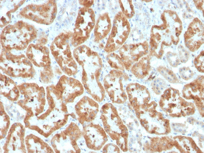
IHC-P analysis of human kidney tissue section using GTX02696 COX2 antibody [COX2/3320R].
![IHC-P analysis of human kidney tissue section using GTX02696 COX2 antibody [COX2/3320R].](https://www.genetex.com/upload/website/prouct_img/normal/GTX02696/GTX02696_20210319_IHC-P_1_w_23053122_461.webp "IHC-P analysis of human kidney tissue section using GTX02696 COX2 antibody [COX2/3320R].")
IHC-P analysis of human kidney tissue section using GTX02696 COX2 antibody [COX2/3320R].
COX2 antibody [COX2/3320R]
GTX02696
ApplicationsImmunoHistoChemistry, ImmunoHistoChemistry Paraffin
Product group Antibodies
ReactivityHuman
TargetPTGS2
Overview
- SupplierGeneTex
- Product NameCOX2 antibody [COX2/3320R]
- Delivery Days Customer9
- Application Supplier NoteIHC-P: 1-2 microg/ml. *Optimal dilutions/concentrations should be determined by the researcher.Not tested in other applications.
- ApplicationsImmunoHistoChemistry, ImmunoHistoChemistry Paraffin
- CertificationResearch Use Only
- ClonalityMonoclonal
- Clone IDCOX2/3320R
- Concentration200 ug/ml
- ConjugateUnconjugated
- Gene ID5743
- Target namePTGS2
- Target descriptionprostaglandin-endoperoxide synthase 2
- Target synonymsCOX-2, COX2, GRIPGHS, PGG/HS, PGHS-2, PHS-2, hCox-2, prostaglandin G/H synthase 2, PGH synthase 2, PHS II, cyclooxygenase 2, cyclooxygenase 2b, prostaglandin H2 synthase 2, prostaglandin-endoperoxide synthase 2 (prostaglandin G/H synthase and cyclooxygenase)
- HostRabbit
- IsotypeIgG
- Protein IDP35354
- Protein NameProstaglandin G/H synthase 2
- Scientific DescriptionProstaglandin-endoperoxide synthase (PTGS), also known as cyclooxygenase, is the key enzyme in prostaglandin biosynthesis, and acts both as a dioxygenase and as a peroxidase. There are two isozymes of PTGS: a constitutive PTGS1 and an inducible PTGS2, which differ in their regulation of expression and tissue distribution. This gene encodes the inducible isozyme. It is regulated by specific stimulatory events, suggesting that it is responsible for the prostanoid biosynthesis involved in inflammation and mitogenesis. [provided by RefSeq, Feb 2009]
- ReactivityHuman
- Storage Instruction2°C to 8°C
- UNSPSC41116161
Datasheet
Related products
Product group Antibodies
Anti-Cox2 AntibodyA35580
ApplicationsImmunoFluorescence, Western Blot, ImmunoHistoChemistry
ReactivityHuman, Mouse, Rat
- SizePrice
Product group Antibodies
Anti-Cox2 Antibody144-62360
ApplicationsImmunoFluorescence, ImmunoPrecipitation, Western Blot, ImmunoHistoChemistry
ReactivityHuman, Mouse, Rat
TargetPTGS2
- SizePrice

Product group Antibodies
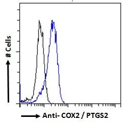
Anti-COX2/Cyclooxygenase 2/PTGS2 Picoband(r) AntibodyA00084-2-CARRIER-FREE
ApplicationsFlow Cytometry, Western Blot, ELISA, ImmunoHistoChemistry
ReactivityHuman, Mouse
TargetPTGS2
- SizePrice

Product group Antibodies
References
Cyclooxygenase 2 Polyclonal AntibodyBS-10411R
ApplicationsImmunoFluorescence, Western Blot, ELISA, ImmunoCytoChemistry, ImmunoHistoChemistry, ImmunoHistoChemistry Frozen, ImmunoHistoChemistry Paraffin
TargetPTGS2
- SizePrice
Product group Antibodies
PTGS2 Monoclonal AntibodyCSB-MA000320
ApplicationsELISA, ImmunoHistoChemistry
ReactivityHuman, Mouse, Rat
TargetPTGS2
- SizePrice
Product group Antibodies
References

Goat anti-COX2 / PTGS2EB05286
ApplicationsFlow Cytometry, ImmunoFluorescence, Western Blot, ELISA
ReactivityBovine, Canine, Human, Mouse, Porcine
TargetPTGS2
- SizePrice
Product group Antibodies
PTGS2 Polyclonal AntibodyCAC15817
ApplicationsWestern Blot, ELISA
TargetPTGS2
- SizePrice
Product group Antibodies
COX2 antibody, C-termGTX15839
ApplicationsFlow Cytometry, ImmunoFluorescence, Western Blot, ImmunoCytoChemistry
ReactivityHuman, Mouse
TargetPTGS2
- SizePrice
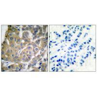
![IHC-P analysis of human prostate carcinoma section using GTX02695 COX2 antibody [COX2/3232R].](https://www.genetex.com/upload/website/prouct_img/normal/GTX02695/GTX02695_20210319_IHC-P_w_23053122_979.webp)
Product group Antibodies
COX2 antibody [COX2/3232R]GTX02695
ApplicationsImmunoHistoChemistry, ImmunoHistoChemistry Paraffin
ReactivityHuman
TargetPTGS2
- SizePrice
![Untreated (–) and treated (+) THP-1 whole cell extract (30 μg) were separated by 7.5% SDS-PAGE, and the membrane was blotted with COX2 antibody [C3], C-term (GTX100656) diluted at 1:500. The HRP-conjugated anti-rabbit IgG antibody (GTX213110-01) was used to detect the primary antibody, and the signal was developed with Trident ECL plus-Enhanced.](https://www.genetex.com/upload/website/prouct_img/normal/GTX100656/GTX100656_43222_20230203_WB_treatment_PMA_LPS_23020621_417.webp)
Product group Antibodies
COX2 antibody [C3], C-termGTX100656
ApplicationsWestern Blot, ImmunoHistoChemistry, ImmunoHistoChemistry Paraffin
ReactivityHuman, Mouse, Rat
TargetPTGS2
- SizePrice