

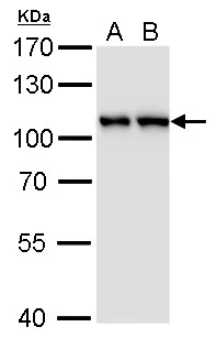
CSE1L antibody [GT5111] detects CSE1L protein by western blot analysis. A. 30 μg PC-12 whole cell lysate/extract B. 30 μg Rat2 whole cell lysate/extract 7.5 % SDS-PAGE CSE1L antibody [GT5111] (GTX630394) dilution: 1:1000
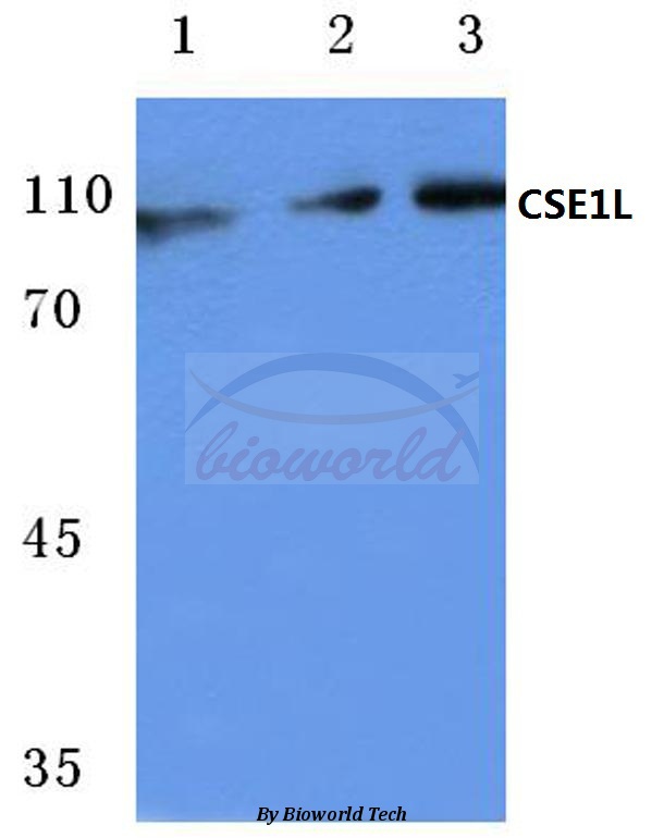
![CSE1L antibody [GT5111] detects CSE1L protein by western blot analysis. A. 30 μg 293T whole cell lysate/extract B. 30 μg A431 whole cell lysate/extract C. 30 μg HeLa whole cell lysate/extract D. 30 μg HepG2 whole cell lysate/extract 7.5 % SDS-PAGE CSE1L antibody [GT5111] (GTX630394) dilution: 1:1000](https://www.genetex.com/upload/website/prouct_img/normal/GTX630394/GTX630394_41526_WB_w_23061202_698.webp "CSE1L antibody [GT5111] detects CSE1L protein by western blot analysis. A. 30 μg 293T whole cell lysate/extract B. 30 μg A431 whole cell lysate/extract C. 30 μg HeLa whole cell lysate/extract D. 30 μg HepG2 whole cell lysate/extract 7.5 % SDS-PAGE CSE1L antibody [GT5111] (GTX630394) dilution: 1:1000")
![CSE1L antibody [GT5111] detects CSE1L protein by western blot analysis. A. 30 μg Neuro2A whole cell lysate/extract B. 30 μg GL261 whole cell lysate/extract C. 30 μg C8D30 whole cell lysate/extract D. 30 μg NIH-3T3 whole cell lysate/extrac E. 30 μg BCL-1 whole cell lysate/extrac F. 30 μg Raw 264.7 whole cell lysate/extract G. 30 μg C2Cl2 whole cell lysate/extract 7.5 % SDS-PAGE CSE1L antibody [GT5111] (GTX630394) dilution: 1:1000](https://www.genetex.com/upload/website/prouct_img/normal/GTX630394/GTX630394_41526_WB_M_w_23061202_662.webp "CSE1L antibody [GT5111] detects CSE1L protein by western blot analysis. A. 30 μg Neuro2A whole cell lysate/extract B. 30 μg GL261 whole cell lysate/extract C. 30 μg C8D30 whole cell lysate/extract D. 30 μg NIH-3T3 whole cell lysate/extrac E. 30 μg BCL-1 whole cell lysate/extrac F. 30 μg Raw 264.7 whole cell lysate/extract G. 30 μg C2Cl2 whole cell lysate/extract 7.5 % SDS-PAGE CSE1L antibody [GT5111] (GTX630394) dilution: 1:1000")
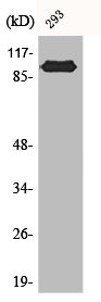
![Non-transfected (–) and transfected (+) 293T whole cell extracts (15 μg) were separated by 7.5% SDS-PAGE, and the membrane was blotted with CSE1L antibody [GT5111] (GTX630394) diluted at 1:1000.](https://www.genetex.com/upload/website/prouct_img/normal/GTX630394/GTX630394_41526_20161103_WB_shRNA_watermark_w_23061202_173.webp "Non-transfected (–) and transfected (+) 293T whole cell extracts (15 μg) were separated by 7.5% SDS-PAGE, and the membrane was blotted with CSE1L antibody [GT5111] (GTX630394) diluted at 1:1000.")
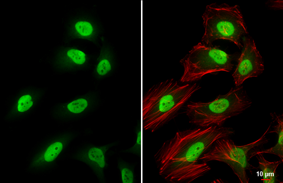
![CSE1L antibody [GT5111] detects CSE1L protein at nucleus by immunofluorescent analysis. Sample: HeLa cells were fixed in 4% paraformaldehyde at RT for 15 min. Green: CSE1L stained by CSE1L antibody [GT5111] (GTX630394) diluted at 1:500. Red: phalloidin, a cytoskeleton marker, diluted at 1:100. Scale bar= 10μm.](https://www.genetex.com/upload/website/prouct_img/normal/GTX630394/GTX630394_41526_20180321_ICC_IF_w_23061202_457.webp "CSE1L antibody [GT5111] detects CSE1L protein at nucleus by immunofluorescent analysis. Sample: HeLa cells were fixed in 4% paraformaldehyde at RT for 15 min. Green: CSE1L stained by CSE1L antibody [GT5111] (GTX630394) diluted at 1:500. Red: phalloidin, a cytoskeleton marker, diluted at 1:100. Scale bar= 10μm.")
CSE1L antibody [GT5111] detects CSE1L protein by western blot analysis. A. 30 μg PC-12 whole cell lysate/extract B. 30 μg Rat2 whole cell lysate/extract 7.5 % SDS-PAGE CSE1L antibody [GT5111] (GTX630394) dilution: 1:1000
CSE1L antibody [GT5111]
GTX630394
ApplicationsImmunoFluorescence, Western Blot, ImmunoCytoChemistry
Product group Antibodies
ReactivityHuman, Mouse, Rat
TargetCSE1L
Overview
- SupplierGeneTex
- Product NameCSE1L antibody [GT5111]
- Delivery Days Customer9
- Application Supplier NoteWB: 1:500-1:3000. ICC/IF: 1:100-1:1000. *Optimal dilutions/concentrations should be determined by the researcher.Not tested in other applications.
- ApplicationsImmunoFluorescence, Western Blot, ImmunoCytoChemistry
- CertificationResearch Use Only
- ClonalityMonoclonal
- Clone IDGT5111
- Concentration1 mg/ml
- ConjugateUnconjugated
- Gene ID1434
- Target nameCSE1L
- Target descriptionchromosome segregation 1 like
- Target synonymsCAS, CSE1, XPO2, exportin-2, BCR/ABL fusion protein, BCR/ABL1 fusion, CSE1 chromosome segregation 1-like, cellular apoptosis susceptibility protein, chromosome segregation 1-like protein, epididymis secretory sperm binding protein, exp2, importin-alpha re-exporter
- HostMouse
- IsotypeIgG2b
- Protein IDP55060
- Protein NameExportin-2
- Scientific DescriptionProteins that carry a nuclear localization signal (NLS) are transported into the nucleus by the importin-alpha/beta heterodimer. Importin-alpha binds the NLS, while importin-beta mediates translocation through the nuclear pore complex. After translocation, RanGTP binds importin-beta and displaces importin-alpha. Importin-alpha must then be returned to the cytoplasm, leaving the NLS protein behind. The protein encoded by this gene binds strongly to NLS-free importin-alpha, and this binding is released in the cytoplasm by the combined action of RANBP1 and RANGAP1. In addition, the encoded protein may play a role both in apoptosis and in cell proliferation. [provided by RefSeq]
- ReactivityHuman, Mouse, Rat
- Storage Instruction-20°C or -80°C,2°C to 8°C
- UNSPSC41116161
Datasheet
Related products
Product group Antibodies
CSE1L AntibodyCSB-PA001218
ApplicationsImmunoFluorescence, ImmunoPrecipitation, Western Blot, ELISA, ImmunoHistoChemistry
ReactivityHuman
TargetCSE1L
- SizePrice
Product group Antibodies

ApplicationsImmunoFluorescence, Western Blot, ELISA, ImmunoCytoChemistry, ImmunoHistoChemistry
- SizePrice
Product group Antibodies
Anti-CSE1L (E2) AntibodyA25109
ApplicationsImmunoFluorescence, ImmunoPrecipitation, Western Blot, ImmunoHistoChemistry
ReactivityHuman, Mouse, Rat
- SizePrice

Product group Antibodies
ApplicationsFlow Cytometry, ImmunoFluorescence, Western Blot, ImmunoCytoChemistry, ImmunoHistoChemistry
ReactivityHuman, Mouse
TargetCSE1L
- SizePrice

Product group Antibodies
Anti-CSE1L AntibodyHPA038059
ApplicationsWestern Blot, ImmunoCytoChemistry, ImmunoHistoChemistry
ReactivityHuman
TargetCSE1L
- SizePrice
Product group Antibodies

CSE1L AntibodyLS-C411044
ApplicationsWestern Blot
ReactivityHuman
TargetCSE1L
- SizePrice
Product group Antibodies
Exportin-2 Recombinant Antibody, AbBy Fluor-405 ConjugatedBSM-61865R-BF405
ApplicationsFlow Cytometry, ImmunoFluorescence, Western Blot
ReactivityHuman, Mouse
TargetCSE1L
- SizePrice
Product group Antibodies
CSE1L Polyclonal AntibodyCAC15773
ApplicationsImmunoFluorescence, Western Blot, ELISA, ImmunoHistoChemistry
TargetCSE1L
- SizePrice
Product group Antibodies
CSE1L antibodyGTX103005
ApplicationsImmunoFluorescence, ImmunoPrecipitation, Western Blot, ImmunoCytoChemistry, ImmunoHistoChemistry, ImmunoHistoChemistry Paraffin
ReactivityHuman, Mouse, Rat
TargetCSE1L
- SizePrice
![CSE1L antibody [GT729] detects CSE1L protein by western blot analysis. A. 30 μg Neuro2A whole cell lysate/extract B. 30 μg GL261 whole cell lysate/extract C. 30 μg C8D30 whole cell lysate/extract D. 30 μg NIH-3T3 whole cell lysate/extrac E. 30 μg BCL-1 whole cell lysate/extrac F. 30 μg Raw 264.7 whole cell lysate/extract G. 30 μg C2Cl2 whole cell lysate/extract 7.5 % SDS-PAGE CSE1L antibody [GT729] (GTX630395) dilution: 1:1000](https://www.genetex.com/upload/website/prouct_img/normal/GTX630395/GTX630395_41526_WB_M_w_23061202_500.webp)
Product group Antibodies
CSE1L antibody [GT729]GTX630395
ApplicationsImmunoFluorescence, Western Blot, ImmunoCytoChemistry
ReactivityHuman, Mouse, Rat
TargetCSE1L
- SizePrice