

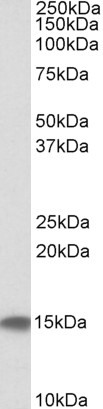
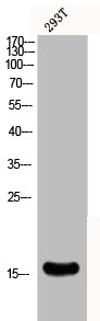
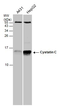
CST3 Antibody Pair
CSB-EAP06197
ApplicationsELISA
Product group Antibodies
ReactivityHuman, Rabbit
TargetCST3
Overview
- SupplierCusabio
- Product NameCST3 Antibody Pair
- Delivery Days Customer9
- Application Supplier NoteWe recommend using the capture antibody at a concentration of 0.35ug/ml and the detection antibody at a concentration of 0.5ug/ml.Optimal dilutions should be determined experimentally by the researcher.
- ApplicationsELISA
- CertificationResearch Use Only
- ConjugateBiotin
- Gene ID1471
- Target nameCST3
- Target descriptioncystatin C
- Target synonymsADLDWA, ARMD11, HEL-S-2, cystatin-C, bA218C14.4 (cystatin C), cystatin 3, epididymis secretory protein Li 2, gamma-trace, neuroendocrine basic polypeptide, post-gamma-globulin
- HostGoat
- IsotypeIgG, IgG2b
- Protein IDP01034
- Protein NameCystatin-C
- ReactivityHuman, Rabbit
- UNSPSC41116163
Related products
Product group Antibodies
Anti-Cystatin C AntibodyA84661
ApplicationsWestern Blot, ELISA
ReactivityHuman
- SizePrice
Product group Antibodies

Anti-Cystatin C [LC01], Human IgG1, kappaAB04698-10.0
ApplicationsELISA
ReactivityHuman
TargetCST3
- SizePrice

Product group Antibodies
Anti-Cystatin C/CST3 Antibody Picoband(r)A00961-2-CARRIER-FREE
ApplicationsWestern Blot, ELISA, ImmunoHistoChemistry
ReactivityHuman, Mouse
TargetCST3
- SizePrice
Product group Antibodies
Anti-CST3 Antibody144-01561
ApplicationsImmunoFluorescence, Western Blot
ReactivityHuman, Mouse, Rat
TargetCST3
- SizePrice
Product group Antibodies
Cystatin-C Recombinant Antibody, AbBy Fluor-350 ConjugatedBSM-61363R-BF350
ApplicationsImmunoFluorescence, Western Blot
ReactivityHuman, Mouse, Rat
TargetCST3
- SizePrice
Product group Antibodies
CST3 AntibodyCSB-PA002010
ApplicationsWestern Blot, ELISA
ReactivityHuman, Monkey, Mouse, Rat
TargetCST3
- SizePrice
Product group Antibodies

Goat anti-CST3 / cystatin CEB09287
ApplicationsWestern Blot, ELISA
ReactivityHuman
TargetCST3
- SizePrice
Product group Antibodies
Cst3 Polyclonal AntibodyCAC07532
ApplicationsWestern Blot, ELISA, ImmunoHistoChemistry
TargetCST3
- SizePrice
Product group Antibodies
Cystatin C antibodyGTX132567
ApplicationsWestern Blot, ImmunoHistoChemistry, ImmunoHistoChemistry Paraffin
ReactivityHuman
TargetCST3
- SizePrice