

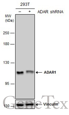
Non-transfected (–) and transfected (+) 293T whole cell extracts (30 μg) were separated by 5% SDS-PAGE, and the membrane was blotted with ADAR1 antibody [GT1066] (GTX629000) diluted at 1:500.
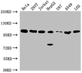
![Various whole cell extracts (30 μg) were separated by 5% SDS-PAGE, and the membrane was blotted with ADAR1 antibody [GT1066] (GTX629000) diluted at 1:1000. The HRP-conjugated anti-mouse IgG antibody (GTX213111-01) was used to detect the primary antibody.](https://www.genetex.com/upload/website/prouct_img/normal/GTX629000/GTX629000_44503_20211112_WB_w_23061202_289.webp "Various whole cell extracts (30 μg) were separated by 5% SDS-PAGE, and the membrane was blotted with ADAR1 antibody [GT1066] (GTX629000) diluted at 1:1000. The HRP-conjugated anti-mouse IgG antibody (GTX213111-01) was used to detect the primary antibody.")
![Immunoprecipitation of ADAR1 protein from 293T whole cell extracts using 5 μg of ADAR1 antibody [GT1066] (GTX629000). Western blot analysis was performed using ADAR1 antibody [GT1066] (GTX629000). EasyBlot anti-Mouse IgG (GTX221667-01) was used as a secondary reagent.](https://www.genetex.com/upload/website/prouct_img/normal/GTX629000/GTX629000_41288_20150612_IP_w_23061202_753.webp "Immunoprecipitation of ADAR1 protein from 293T whole cell extracts using 5 μg of ADAR1 antibody [GT1066] (GTX629000). Western blot analysis was performed using ADAR1 antibody [GT1066] (GTX629000). EasyBlot anti-Mouse IgG (GTX221667-01) was used as a secondary reagent.")
![ADAR1 antibody [GT1066] detects ADAR1 protein at nucleus by immunofluorescent analysis. Sample: HeLa cells were fixed in 4% paraformaldehyde for 10 min. Green: ADAR1 protein stained by ADAR1 antibody [GT1066] (GTX629000) diluted at 1:100. Red: phalloidin, a cytoskeleton marker, diluted at 1:100. Scale bar = 10 μm.](https://www.genetex.com/upload/website/prouct_img/normal/GTX629000/GTX629000_41288_IFA_w_23061202_642.webp "ADAR1 antibody [GT1066] detects ADAR1 protein at nucleus by immunofluorescent analysis. Sample: HeLa cells were fixed in 4% paraformaldehyde for 10 min. Green: ADAR1 protein stained by ADAR1 antibody [GT1066] (GTX629000) diluted at 1:100. Red: phalloidin, a cytoskeleton marker, diluted at 1:100. Scale bar = 10 μm.")
Non-transfected (–) and transfected (+) 293T whole cell extracts (30 μg) were separated by 5% SDS-PAGE, and the membrane was blotted with ADAR1 antibody [GT1066] (GTX629000) diluted at 1:500.
ADAR1 antibody [GT1066]
GTX629000
ApplicationsImmunoFluorescence, ImmunoPrecipitation, Western Blot, ImmunoCytoChemistry
Product group Antibodies
ReactivityHuman
TargetADAR
Overview
- SupplierGeneTex
- Product NameADAR1 antibody [GT1066]
- Delivery Days Customer9
- Application Supplier NoteWB: 1:500-1:3000. ICC/IF: 1:100-1:1000. IP: 1:100-1:500. *Optimal dilutions/concentrations should be determined by the researcher.Not tested in other applications.
- ApplicationsImmunoFluorescence, ImmunoPrecipitation, Western Blot, ImmunoCytoChemistry
- CertificationResearch Use Only
- ClonalityMonoclonal
- Clone IDGT1066
- Concentration1 mg/ml
- ConjugateUnconjugated
- Gene ID103
- Target nameADAR
- Target descriptionadenosine deaminase RNA specific
- Target synonymsADAR1, AGS6, DRADA, DSH, DSRAD, G1P1, IFI-4, IFI4, K88DSRBP, P136, double-stranded RNA-specific adenosine deaminase, 136 kDa double-stranded RNA-binding protein, adenosine deaminase acting on RNA 1-A, dsRNA adenosine deaminase, dsRNA adeonosine deaminase, interferon-induced protein 4, interferon-inducible protein 4
- HostMouse
- IsotypeIgG1
- Protein IDP55265
- Protein NameDouble-stranded RNA-specific adenosine deaminase
- Scientific DescriptionThis gene encodes the enzyme responsible for RNA editing by site-specific deamination of adenosines. This enzyme destabilizes double stranded RNA through conversion of adenosine to inosine. Mutations in this gene have been associated with dyschromatosis symmetrica hereditaria. Alternate transcriptional splice variants, encoding different isoforms, have been characterized. [provided by RefSeq]
- ReactivityHuman
- Storage Instruction-20°C or -80°C,2°C to 8°C
- UNSPSC41116161
Datasheet
Related products
Product group Antibodies
Anti-ADAR1 AntibodyA97053
ApplicationsELISA, ImmunoHistoChemistry
ReactivityHuman, Mouse, Rat
- SizePrice

Product group Antibodies
Anti-ADAR1/ADAR Antibody Picoband(r)A00869-2-CARRIER-FREE
ApplicationsImmunoFluorescence, Western Blot, ELISA, ImmunoCytoChemistry, ImmunoHistoChemistry
ReactivityHuman, Mouse, Rat
TargetADAR
- SizePrice
Product group Antibodies
Anti-ADAR Antibody144-07869
ApplicationsImmunoFluorescence, Western Blot, ImmunoHistoChemistry
ReactivityHuman, Mouse, Rat
TargetADAR
- SizePrice

Product group Antibodies
Anti-ADAR AntibodyAMAB90535
ApplicationsWestern Blot, ImmunoCytoChemistry, ImmunoHistoChemistry
ReactivityHuman
TargetADAR
- SizePrice
Product group Antibodies
ADAR1 Polyclonal AntibodyBS-2168R
ApplicationsImmunoFluorescence, Western Blot, ELISA, ImmunoCytoChemistry, ImmunoHistoChemistry, ImmunoHistoChemistry Frozen, ImmunoHistoChemistry Paraffin
ReactivityHuman, Mouse, Rat
TargetADAR
- SizePrice
Product group Antibodies
ADAR AntibodyCSB-PA001324LA01HU
ApplicationsWestern Blot, ELISA, ImmunoHistoChemistry
ReactivityHuman
TargetADAR
- SizePrice
Product group Antibodies
Adar Polyclonal AntibodyCAC11643
ApplicationsWestern Blot, ELISA, ImmunoHistoChemistry
TargetADAR
- SizePrice
![ADAR1 antibody [N3C1], Internal detects ADAR1 protein at nucleolus and nucleus by immunofluorescent analysis. Sample: HeLa cells were fixed in 4% paraformaldehyde at RT for 15 min. Green: ADAR1 protein stained by ADAR1 antibody [N3C1], Internal (GTX101602) diluted at 1:200. Red: phalloidin, a cytoskeleton marker, diluted at 1:200. Blue: Hoechst 33342 staining. Scale bar = 10 μm.](https://www.genetex.com/upload/website/prouct_img/normal/GTX101602/GTX101602_40506_20150714_IFA_w_23060100_395.webp)
Product group Antibodies
ADAR1 antibody [N3C1], InternalGTX101602
ApplicationsImmunoFluorescence, ImmunoPrecipitation, Western Blot, ImmunoCytoChemistry, ImmunoHistoChemistry, ImmunoHistoChemistry Paraffin
ReactivityHuman, Rat
TargetADAR
- SizePrice
![Various whole cell extracts (30 μg) were separated by 5% SDS-PAGE, and the membrane was blotted with ADAR1 antibody [HL1789] (GTX637441) diluted at 1:1000. The HRP-conjugated anti-rabbit IgG antibody (GTX213110-01) was used to detect the primary antibody.](https://www.genetex.com/upload/website/prouct_img/normal/GTX637441/GTX637441_T-44809_20220923_WB_22092622_873.webp)
Product group Antibodies
ADAR1 antibody [HL1789]GTX637441
ApplicationsWestern Blot, ImmunoHistoChemistry, ImmunoHistoChemistry Paraffin
ReactivityHuman
TargetADAR
- SizePrice