

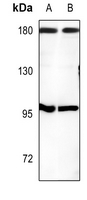
WB analysis of mouse muscle (A), rat muscle (B) tissue lysates using GTX56275 CUTL1 antibody.

")
WB analysis of mouse muscle (A), rat muscle (B) tissue lysates using GTX56275 CUTL1 antibody.
CUTL1 antibody
GTX56275
ApplicationsImmunoFluorescence, Western Blot, ImmunoCytoChemistry, ImmunoHistoChemistry, ImmunoHistoChemistry Paraffin
Product group Antibodies
ReactivityHuman, Mouse, Rat
TargetCUX1
Overview
- SupplierGeneTex
- Product NameCUTL1 antibody
- Delivery Days Customer9
- Application Supplier NoteWB: 1:500 - 1:1000. ICC/IF: 1:100 - 1:500. IHC-P: 1:100 - 1:200. *Optimal dilutions/concentrations should be determined by the researcher.Not tested in other applications.
- ApplicationsImmunoFluorescence, Western Blot, ImmunoCytoChemistry, ImmunoHistoChemistry, ImmunoHistoChemistry Paraffin
- CertificationResearch Use Only
- ClonalityPolyclonal
- ConjugateUnconjugated
- Gene ID1523
- Target nameCUX1
- Target descriptioncut like homeobox 1
- Target synonymsCASP, CDP, CDP/Cut, CDP1, COY1, CUTL1, CUX, Clox, Cux/CDP, GDDI, GOLIM6, NEDDMS, Nbla10317, p100, p110, p200, p75, protein CASP, Homeobox protein cut-like 1, CCAAT displacement protein, CDP/Cux p200, CUX1 gene Alternatively Spliced Product, cut homolog, golgi integral membrane protein 6, homeobox protein cux-1, mutant CUX1, putative protein product of Nbla10317
- HostRabbit
- IsotypeIgG
- Protein IDP39880
- Protein NameHomeobox protein cut-like 1
- Scientific DescriptionThe protein encoded by this gene is a member of the homeodomain family of DNA binding proteins. It may regulate gene expression, morphogenesis, and differentiation and it may also play a role in the cell cycle progession. Several alternatively spliced transcript variants encoding different isoforms have been identified.[provided by RefSeq, Feb 2011]
- ReactivityHuman, Mouse, Rat
- Storage Instruction-20°C or -80°C,2°C to 8°C
- UNSPSC41116161
References
- Histological Analysis of a Mouse Model of the 22q11.2 Microdeletion Syndrome.Read this paper
Datasheet
Related products
Product group Antibodies
Anti-Mouse/Rat CUX1 Antibody144-02213
ApplicationsWestern Blot
ReactivityHuman, Mouse, Rat
TargetCUX1
- SizePrice

Product group Antibodies
Anti-CUX1 AntibodyAMAB91352
ApplicationsImmunoCytoChemistry, ImmunoHistoChemistry
ReactivityHuman
TargetCUX1
- SizePrice
Product group Antibodies
Anti-CUX1 AntibodyA34476
ApplicationsImmunoFluorescence, Western Blot, ELISA, ImmunoHistoChemistry
ReactivityHuman, Mouse
- SizePrice
Product group Antibodies

CUX1 / CASP AntibodyLS-C669861
ApplicationsWestern Blot, ELISA, ImmunoHistoChemistry, ImmunoHistoChemistry Paraffin
ReactivityHuman
TargetCUX1
- SizePrice

Product group Antibodies
Anti-CUX1 Antibody Picoband(r)A01653-4-CARRIER-FREE
ApplicationsFlow Cytometry, Western Blot, ELISA, ImmunoHistoChemistry
ReactivityHuman, Mouse
TargetCUX1
- SizePrice
Product group Antibodies
CUX1/CUX1 AntibodyCSB-PA001549
ApplicationsImmunoFluorescence, Western Blot, ELISA, ImmunoHistoChemistry
ReactivityHuman, Mouse
TargetCUX1
- SizePrice
Product group Antibodies
CUX1 Polyclonal AntibodyCAC15377
ApplicationsWestern Blot, ELISA, ImmunoHistoChemistry
ReactivityMouse, Rat
TargetCUX1
- SizePrice
![CUTL1 antibody detects CUTL1 protein by immunofluorescent analysis. Sample: DIV10 rat E18 primary cortical neuron cells were fixed in 4% paraformaldehyde at RT for 15 min. Green: CUTL1 stained by CUTL1 antibody (GTX114351) diluted at 1:500. Red: Tau, stained by Tau antibody [GT287] (GTX634809) diluted at 1:500. Blue: Fluoroshield with DAPI (GTX30920).](https://www.genetex.com/upload/website/prouct_img/normal/GTX114351/GTX114351_40856_20191125_ICC_IF_R_w_23060501_273.webp)
Product group Antibodies
CUTL1 antibodyGTX114351
ApplicationsImmunoFluorescence, ImmunoPrecipitation, Western Blot, ImmunoCytoChemistry
ReactivityHuman, Rat
TargetCUX1
- SizePrice
Product group Antibodies
Anti-CUX1 AntibodyCAB2213
ApplicationsImmunoFluorescence, Western Blot, ELISA, ImmunoCytoChemistry, ImmunoHistoChemistry, ImmunoHistoChemistry Paraffin
ReactivityHuman
TargetCUX1
- SizePrice