

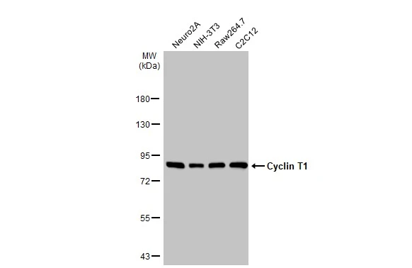
Various whole cell extracts (30 μg) were separated by 7.5% SDS-PAGE, and the membrane was blotted with Cyclin T1 antibody (GTX133413) diluted at 1:1000. The HRP-conjugated anti-rabbit IgG antibody (GTX213110-01) was used to detect the primary antibody.
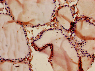
 diluted at 1:500. Red: Phalloidin, a cytoskeleton marker, diluted at 1:100.")
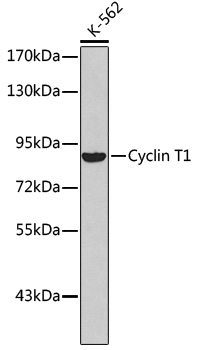
 were separated by 7.5% SDS-PAGE, and the membrane was blotted with Cyclin T1 antibody (GTX133413) diluted at 1:1000. The HRP-conjugated anti-rabbit IgG antibody (GTX213110-01) was used to detect the primary antibody.")
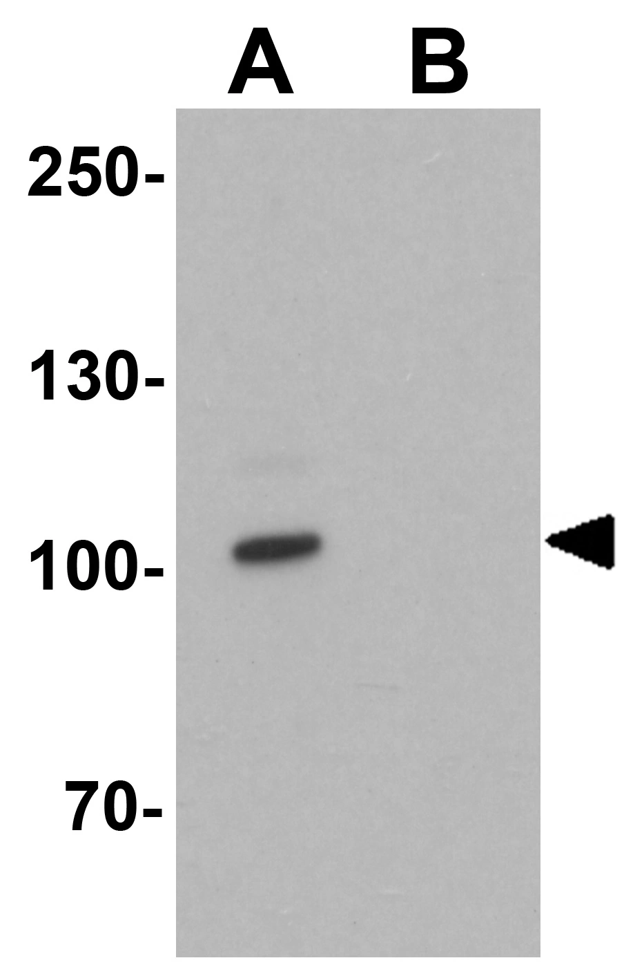
 were separated by 7.5% SDS-PAGE, and the membrane was blotted with Cyclin T1 antibody (GTX133413) diluted at 1:1000.")
Various whole cell extracts (30 μg) were separated by 7.5% SDS-PAGE, and the membrane was blotted with Cyclin T1 antibody (GTX133413) diluted at 1:1000. The HRP-conjugated anti-rabbit IgG antibody (GTX213110-01) was used to detect the primary antibody.
Cyclin T1 antibody
GTX133413
ApplicationsImmunoFluorescence, Western Blot, ImmunoCytoChemistry
Product group Antibodies
ReactivityHuman, Mouse, Rat
TargetCCNT1
Overview
- SupplierGeneTex
- Product NameCyclin T1 antibody
- Delivery Days Customer9
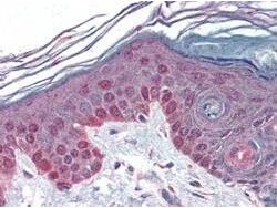
- Application Supplier NoteWB: 1:500-1:3000. ICC/IF: 1:100-1:1000. *Optimal dilutions/concentrations should be determined by the researcher.Not tested in other applications.
- ApplicationsImmunoFluorescence, Western Blot, ImmunoCytoChemistry
- CertificationResearch Use Only
- ClonalityPolyclonal
- Concentration1 mg/ml
- ConjugateUnconjugated
- Gene ID904
- Target nameCCNT1
- Target descriptioncyclin T1
- Target synonymsCCNT, CYCT1, HIVE1, cyclin-T1, CDK9-associated C-type protein, MLLT10/CCNT1 fusion, cyclin C-related protein, human immunodeficiency virus type 1 (HIV-1) expression (elevated) 1
- HostRabbit
- IsotypeIgG
- Protein IDO60563
- Protein NameCyclin-T1
- Scientific DescriptionThe protein encoded by this gene belongs to the highly conserved cyclin family, whose members are characterized by a dramatic periodicity in protein abundance through the cell cycle. Cyclins function as regulators of CDK kinases. Different cyclins exhibit distinct expression and degradation patterns which contribute to the temporal coordination of each mitotic event. This cyclin tightly associates with CDK9 kinase, and was found to be a major subunit of the transcription elongation factor p-TEFb. The kinase complex containing this cyclin and the elongation factor can interact with, and act as a cofactor of human immunodeficiency virus type 1 (HIV-1) Tat protein, and was shown to be both necessary and sufficient for full activation of viral transcription. This cyclin and its kinase partner were also found to be involved in the phosphorylation and regulation of the carboxy-terminal domain (CTD) of the largest RNA polymerase II subunit. [provided by RefSeq]
- ReactivityHuman, Mouse, Rat
- Storage Instruction-20°C or -80°C,2°C to 8°C
- UNSPSC41116161
Datasheet
Related products
Product group Antibodies
CCNT1 AntibodyCSB-PA004832LA01HU
ApplicationsELISA, ImmunoHistoChemistry
ReactivityHuman
TargetCCNT1
- SizePrice
Product group Antibodies
ApplicationsWestern Blot
ReactivityHuman
- SizePrice

Product group Antibodies
Anti-CCNT1 AntibodyHPA004892
ApplicationsWestern Blot, ImmunoCytoChemistry, ImmunoHistoChemistry
ReactivityHuman, Mouse, Rat
TargetCCNT1
- SizePrice
Product group Antibodies

CCNT1 / Cyclin T1 AntibodyLS-C331858
ApplicationsWestern Blot, ImmunoHistoChemistry
ReactivityHuman
TargetCCNT1
- SizePrice

Product group Antibodies
Anti-Cyclin T1/CCNT1 Antibody Picoband(r)PB9990-CARRIER-FREE
ApplicationsFlow Cytometry, ImmunoFluorescence, Western Blot, ImmunoCytoChemistry, ImmunoHistoChemistry, ImmunoHistoChemistry Frozen
ReactivityBovine, Human, Mouse, Rat
TargetCCNT1
- SizePrice
Product group Antibodies
Cyclin T1 antibodyGTX17200
ApplicationsWestern Blot, ELISA, ImmunoHistoChemistry, ImmunoHistoChemistry Paraffin
ReactivityHuman, Mouse, Rat
TargetCCNT1
- SizePrice
Product group Antibodies
Cyclin T1 antibodyGTX22098
ApplicationsImmunoPrecipitation, Western Blot, ELISA, ImmunoHistoChemistry, ImmunoHistoChemistry Paraffin
ReactivityHuman, Mouse, Rat
TargetCCNT1
- SizePrice