

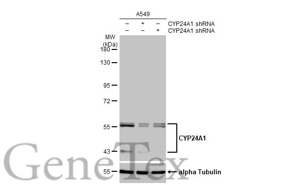
Non-transfected (–) and transfected (+) A549 whole cell extracts (30 μg) were separated by 7.5% SDS-PAGE, and the membrane was blotted with CYP24A1 antibody [HL1783] (GTX637435) diluted at 1:1000. The HRP-conjugated anti-rabbit IgG antibody (GTX213110-01) was used to detect the primary antibody, and the signal was developed with Trident ECL plus-Enhanced.
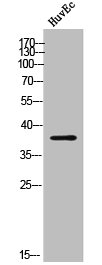
![Various whole cell extracts (30 μg) were separated by 7.5% SDS-PAGE, and the membrane was blotted with CYP24A1 antibody [HL1783] (GTX637435) diluted at 1:2000. The HRP-conjugated anti-rabbit IgG antibody (GTX213110-01) was used to detect the primary antibody. Corresponding RNA expression data for the same cell lines are based on Human Protein Atlas program.](https://www.genetex.com/upload/website/prouct_img/normal/GTX637435/GTX637435_44970_20230317_WB_TPM_watermark_23032022_766.webp "Various whole cell extracts (30 μg) were separated by 7.5% SDS-PAGE, and the membrane was blotted with CYP24A1 antibody [HL1783] (GTX637435) diluted at 1:2000. The HRP-conjugated anti-rabbit IgG antibody (GTX213110-01) was used to detect the primary antibody. Corresponding RNA expression data for the same cell lines are based on Human Protein Atlas program.")
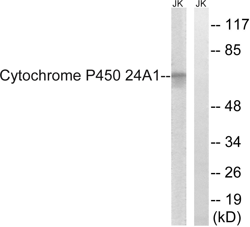
![Various whole cell extracts (30 μg) were separated by 10% SDS-PAGE, and the membrane was blotted with CYP24A1 antibody [HL1783] (GTX637435) diluted at 1:1000. The HRP-conjugated anti-rabbit IgG antibody (GTX213110-01) was used to detect the primary antibody.](https://www.genetex.com/upload/website/prouct_img/normal/GTX637435/GTX637435_44970_20230421_WB_M_R_23042500_398.webp "Various whole cell extracts (30 μg) were separated by 10% SDS-PAGE, and the membrane was blotted with CYP24A1 antibody [HL1783] (GTX637435) diluted at 1:1000. The HRP-conjugated anti-rabbit IgG antibody (GTX213110-01) was used to detect the primary antibody.")
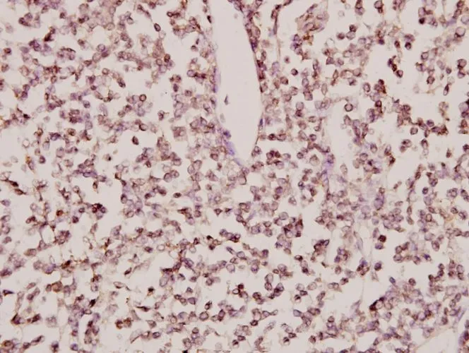
![CYP24A1 antibody [HL1783] detects CYP24A1 protein at mitochondria by immunohistochemical analysis. Sample: Paraffin-embedded human oral carcinoma. CYP24A1 stained by CYP24A1 antibody [HL1783] (GTX637435) diluted at 1:200. Antigen Retrieval: Citrate buffer, pH 6.0, 15 min](https://www.genetex.com/upload/website/prouct_img/normal/GTX637435/GTX637435_44970_20230602_IHC-P_23061320_258.webp "CYP24A1 antibody [HL1783] detects CYP24A1 protein at mitochondria by immunohistochemical analysis. Sample: Paraffin-embedded human oral carcinoma. CYP24A1 stained by CYP24A1 antibody [HL1783] (GTX637435) diluted at 1:200. Antigen Retrieval: Citrate buffer, pH 6.0, 15 min")
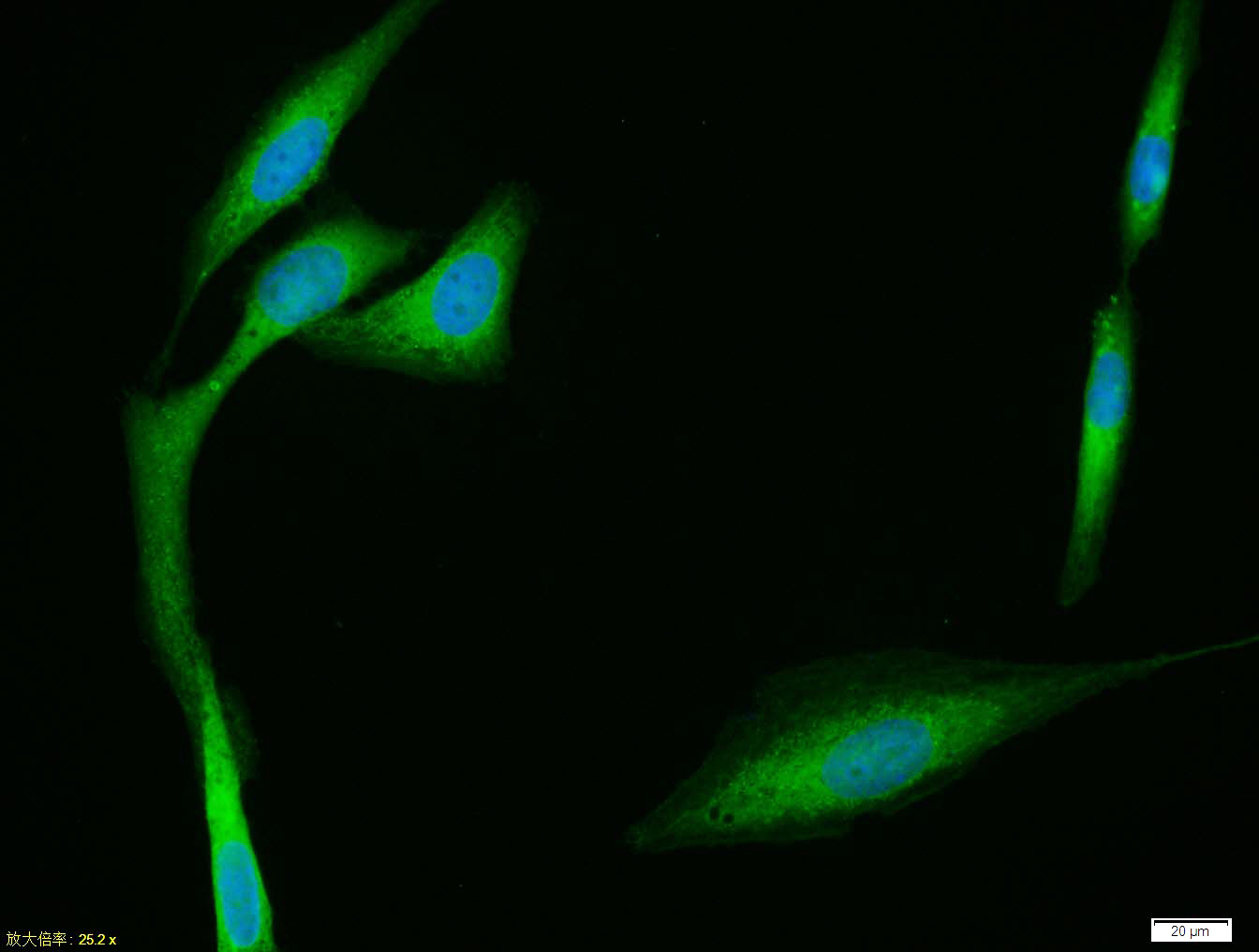
![CYP24A1 antibody [HL1783] detects CYP24A1 protein at mitochondria by immunofluorescent analysis. Sample: A549 cells were fixed in 4% paraformaldehyde at RT for 15 min. Green: CYP24A1 stained by CYP24A1 antibody [HL1783] (GTX637435) diluted at 1:500. Red: alpha Tubulin, a cytoskeleton marker, stained by alpha Tubulin antibody [GT114] (GTX628802) diluted at 1:1000. Blue: Fluoroshield with DAPI (GTX30920).](https://www.genetex.com/upload/website/prouct_img/normal/GTX637435/GTX637435_44970_20230609_ICC_IF_23062718_869.webp "CYP24A1 antibody [HL1783] detects CYP24A1 protein at mitochondria by immunofluorescent analysis. Sample: A549 cells were fixed in 4% paraformaldehyde at RT for 15 min. Green: CYP24A1 stained by CYP24A1 antibody [HL1783] (GTX637435) diluted at 1:500. Red: alpha Tubulin, a cytoskeleton marker, stained by alpha Tubulin antibody [GT114] (GTX628802) diluted at 1:1000. Blue: Fluoroshield with DAPI (GTX30920).")
Non-transfected (–) and transfected (+) A549 whole cell extracts (30 μg) were separated by 7.5% SDS-PAGE, and the membrane was blotted with CYP24A1 antibody [HL1783] (GTX637435) diluted at 1:1000. The HRP-conjugated anti-rabbit IgG antibody (GTX213110-01) was used to detect the primary antibody, and the signal was developed with Trident ECL plus-Enhanced.
CYP24A1 antibody [HL1783]
GTX637435
ApplicationsImmunoFluorescence, Western Blot, ImmunoCytoChemistry, ImmunoHistoChemistry, ImmunoHistoChemistry Paraffin
Product group Antibodies
ReactivityHuman, Mouse, Rat
TargetCYP24A1
Overview
- SupplierGeneTex
- Product NameCYP24A1 antibody [HL1783]
- Delivery Days Customer9
- Application Supplier NoteWB: 1:500-1:3000. *Optimal dilutions/concentrations should be determined by the researcher.Not tested in other applications.
- ApplicationsImmunoFluorescence, Western Blot, ImmunoCytoChemistry, ImmunoHistoChemistry, ImmunoHistoChemistry Paraffin
- CertificationResearch Use Only
- ClonalityMonoclonal
- Clone IDHL1783
- Concentration1 mg/ml
- ConjugateUnconjugated
- Gene ID1591
- Target nameCYP24A1
- Target descriptioncytochrome P450 family 24 subfamily A member 1
- Target synonymsCP24, CYP24, HCAI, HCINF1, P450-CC24, 1,25-dihydroxyvitamin D(3) 24-hydroxylase, mitochondrial, 1,25-@dihydroxyvitamin D3 24-hydroxylase, 24-OHase, cytochrome P450 24A1, cytochrome P450, family 24, subfamily A, polypeptide 1, cytochrome P450, subfamily XXIV (vitamin D 24-hydroxylase), cytochrome P450-CC24, exo-mitochondrial protein, lncBCAS1-4_1, vitamin D 24-hydroxylase, vitamin D(3) 24-hydroxylase
- HostRabbit
- IsotypeIgG
- Protein IDQ07973
- Protein Name1,25-dihydroxyvitamin D(3) 24-hydroxylase, mitochondrial
- Scientific DescriptionThis gene encodes a member of the cytochrome P450 superfamily of enzymes. The cytochrome P450 proteins are monooxygenases which catalyze many reactions involved in drug metabolism and synthesis of cholesterol, steroids and other lipids. This mitochondrial protein initiates the degradation of 1,25-dihydroxyvitamin D3, the physiologically active form of vitamin D3, by hydroxylation of the side chain. In regulating the level of vitamin D3, this enzyme plays a role in calcium homeostasis and the vitamin D endocrine system. Alternatively spliced transcript variants encoding different isoforms have been found for this gene. [provided by RefSeq, Jul 2008]
- ReactivityHuman, Mouse, Rat
- Storage Instruction-20°C or -80°C,2°C to 8°C
- UNSPSC41116161
Datasheet
Related products
Product group Antibodies
CYP24A1 AntibodyCSB-PA007611
ApplicationsWestern Blot, ELISA, ImmunoHistoChemistry
ReactivityHuman
TargetCYP24A1
- SizePrice
Product group Antibodies
ApplicationsWestern Blot, ELISA
ReactivityHuman
- SizePrice
Product group Antibodies

Goat anti-CYP24A1EB10490
ApplicationsWestern Blot, ELISA, ImmunoHistoChemistry
ReactivityHuman, Rat
TargetCYP24A1
- SizePrice

Product group Antibodies
Anti-CYP24A1 AntibodyHPA022261
ApplicationsWestern Blot, ImmunoCytoChemistry, ImmunoHistoChemistry
ReactivityHuman
TargetCYP24A1
- SizePrice
Product group Antibodies

CYP24 / CYP24A1 Antibody (C-Terminus)LS-C368862
ApplicationsWestern Blot, ImmunoHistoChemistry, ImmunoHistoChemistry Paraffin
ReactivityHuman, Monkey
TargetCYP24A1
- SizePrice
Product group Antibodies
ApplicationsImmunoPrecipitation, Western Blot, ImmunoCytoChemistry, ImmunoHistoChemistry
ReactivityMouse, Rat
TargetCYP24A1
- SizePrice

Product group Antibodies
Anti-CYP24A1 Antibody Picoband(r)PB9547-CARRIER-FREE
ApplicationsWestern Blot, ImmunoHistoChemistry
ReactivityHuman, Mouse, Rat
TargetCYP24A1
- SizePrice
Product group Antibodies
CYP24A1 Polyclonal AntibodyBS-3899R
ApplicationsImmunoFluorescence, ELISA, ImmunoCytoChemistry, ImmunoHistoChemistry, ImmunoHistoChemistry Frozen, ImmunoHistoChemistry Paraffin
ReactivityBovine, Canine, Chicken, Equine, Human, Mouse, Porcine, Rabbit, Rat
TargetCYP24A1
- SizePrice
Product group Antibodies
CYP24A1 antibodyGTX66663
ApplicationsImmunoHistoChemistry, ImmunoHistoChemistry Paraffin
ReactivityHuman
TargetCYP24A1
- SizePrice
Product group Antibodies
Anti-CYP24A1 Antibody144-65277
ApplicationsImmunoFluorescence, ImmunoHistoChemistry
ReactivityHuman, Mouse, Rat
TargetCYP24A1
- SizePrice