

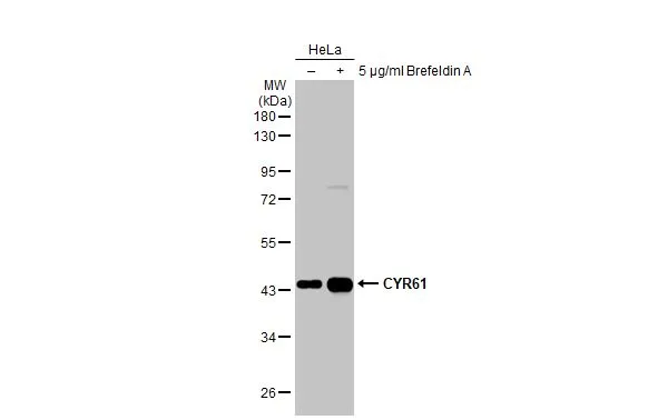
Untreated (–) and treated (+) HeLa whole cell extracts (30 μg) were separated by 10% SDS-PAGE, and the membrane was blotted with CYR61 antibody [N1C3] (GTX103669) diluted at 1:1000. The HRP-conjugated anti-rabbit IgG antibody (GTX213110-01) was used to detect the primary antibody.
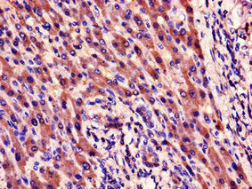
![CYR61 antibody [N1C3] detects secreted CYR61 protein by immunohistochemical analysis. Sample: Paraffin-embedded human colon cancer. CYR61 stained by CYR61 antibody [N1C3] (GTX103669) diluted at 1:500. Antigen Retrieval: Citrate buffer, pH 6.0, 15 min](https://www.genetex.com/upload/website/prouct_img/normal/GTX103669/GTX103669_44622_20221028_IHC-P_22122018_892.webp "CYR61 antibody [N1C3] detects secreted CYR61 protein by immunohistochemical analysis. Sample: Paraffin-embedded human colon cancer. CYR61 stained by CYR61 antibody [N1C3] (GTX103669) diluted at 1:500. Antigen Retrieval: Citrate buffer, pH 6.0, 15 min")
![CYR61 antibody [N1C3] detects secreted CYR61 protein by immunohistochemical analysis. Sample: Paraffin-embedded rat colon. CYR61 stained by CYR61 antibody [N1C3] (GTX103669) diluted at 1:500. Antigen Retrieval: Citrate buffer, pH 6.0, 15 min](https://www.genetex.com/upload/website/prouct_img/normal/GTX103669/GTX103669_44622_20220520_IHC-P_R_w_23060119_514.webp "CYR61 antibody [N1C3] detects secreted CYR61 protein by immunohistochemical analysis. Sample: Paraffin-embedded rat colon. CYR61 stained by CYR61 antibody [N1C3] (GTX103669) diluted at 1:500. Antigen Retrieval: Citrate buffer, pH 6.0, 15 min")
![CYR61 antibody [N1C3] detects CYR61 protein at cytoplasm in rat intestine by immunohistochemical analysis. Sample: Paraffin-embedded rat intestine. CYR61 antibody [N1C3] (GTX103669) diluted at 1:500.
Antigen Retrieval: Citrate buffer, pH 6.0, 15 min](https://www.genetex.com/upload/website/prouct_img/normal/GTX103669/GTX103669_40150_20151030_IHC-P_R_w_23060119_153.webp "CYR61 antibody [N1C3] detects CYR61 protein at cytoplasm in rat intestine by immunohistochemical analysis. Sample: Paraffin-embedded rat intestine. CYR61 antibody [N1C3] (GTX103669) diluted at 1:500.
Antigen Retrieval: Citrate buffer, pH 6.0, 15 min")
![CYR61 antibody [N1C3] detects CYR61 protein at endoplasmic reticulum by immunofluorescent analysis. Sample: HeLa cells were fixed in 4% paraformaldehyde at RT for 15 min. Green: CYR61 protein stained by CYR61 antibody [N1C3] (GTX103669) diluted at 1:200. Blue: Hoechst 33342 staining.](https://www.genetex.com/upload/website/prouct_img/normal/GTX103669/GTX103669_40150_20150924_IFA_w_23060119_339.webp "CYR61 antibody [N1C3] detects CYR61 protein at endoplasmic reticulum by immunofluorescent analysis. Sample: HeLa cells were fixed in 4% paraformaldehyde at RT for 15 min. Green: CYR61 protein stained by CYR61 antibody [N1C3] (GTX103669) diluted at 1:200. Blue: Hoechst 33342 staining.")
![CYR61 antibody [N1C3] detects secreted CYR61 protein by immunohistochemical analysis. Sample: Paraffin-embedded mouse intestine. CYR61 stained by CYR61 antibody [N1C3] (GTX103669) diluted at 1:500. Antigen Retrieval: Citrate buffer, pH 6.0, 15 min](https://www.genetex.com/upload/website/prouct_img/normal/GTX103669/GTX103669_44622_20220422_IHC-P_M_w_23060119_367.webp "CYR61 antibody [N1C3] detects secreted CYR61 protein by immunohistochemical analysis. Sample: Paraffin-embedded mouse intestine. CYR61 stained by CYR61 antibody [N1C3] (GTX103669) diluted at 1:500. Antigen Retrieval: Citrate buffer, pH 6.0, 15 min")
![CYR61 antibody [N1C3] detects secreted CYR61 protein by immunohistochemical analysis. Sample: Paraffin-embedded mouse colon. CYR61 stained by CYR61 antibody [N1C3] (GTX103669) diluted at 1:500. Antigen Retrieval: Citrate buffer, pH 6.0, 15 min](https://www.genetex.com/upload/website/prouct_img/normal/GTX103669/GTX103669_40150_20220318_IHC-P_M_w_23060119_373.webp "CYR61 antibody [N1C3] detects secreted CYR61 protein by immunohistochemical analysis. Sample: Paraffin-embedded mouse colon. CYR61 stained by CYR61 antibody [N1C3] (GTX103669) diluted at 1:500. Antigen Retrieval: Citrate buffer, pH 6.0, 15 min")
![CYR61 antibody [N1C3] detects secreted CYR61 protein by immunohistochemical analysis. Sample: Paraffin-embedded rat colon. CYR61 stained by CYR61 antibody [N1C3] (GTX103669) diluted at 1:500. Antigen Retrieval: Citrate buffer, pH 6.0, 15 min](https://www.genetex.com/upload/website/prouct_img/normal/GTX103669/GTX103669_40150_20220318_IHC-P_R_w_23060119_881.webp "CYR61 antibody [N1C3] detects secreted CYR61 protein by immunohistochemical analysis. Sample: Paraffin-embedded rat colon. CYR61 stained by CYR61 antibody [N1C3] (GTX103669) diluted at 1:500. Antigen Retrieval: Citrate buffer, pH 6.0, 15 min")
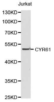
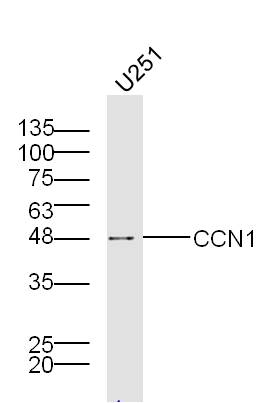
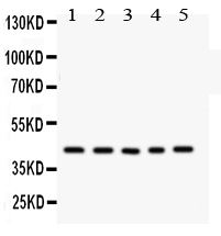
![Various whole cell extracts (30 μg) were separated by 10% SDS-PAGE, and the membrane was blotted with CYR61 antibody [N1C3] (GTX103669) diluted at 1:1000. The HRP-conjugated anti-rabbit IgG antibody (GTX213110-01) was used to detect the primary antibody.](https://www.genetex.com/upload/website/prouct_img/normal/GTX103669/GTX103669_44853_20221104_WB_24011618_939.webp "Various whole cell extracts (30 μg) were separated by 10% SDS-PAGE, and the membrane was blotted with CYR61 antibody [N1C3] (GTX103669) diluted at 1:1000. The HRP-conjugated anti-rabbit IgG antibody (GTX213110-01) was used to detect the primary antibody.")
Untreated (–) and treated (+) HeLa whole cell extracts (30 μg) were separated by 10% SDS-PAGE, and the membrane was blotted with CYR61 antibody [N1C3] (GTX103669) diluted at 1:1000. The HRP-conjugated anti-rabbit IgG antibody (GTX213110-01) was used to detect the primary antibody.
CYR61 antibody [N1C3]
GTX103669
ApplicationsImmunoFluorescence, Western Blot, ImmunoCytoChemistry, ImmunoHistoChemistry, ImmunoHistoChemistry Paraffin
Product group Antibodies
ReactivityHuman, Mouse, Rat
TargetCCN1
Overview
- SupplierGeneTex
- Product NameCYR61 antibody [N1C3]
- Delivery Days Customer9
- Application Supplier NoteWB: 1:500-1:3000. ICC/IF: 1:100-1:1000. IHC-P: 1:100-1:1000. *Optimal dilutions/concentrations should be determined by the researcher.Not tested in other applications.
- ApplicationsImmunoFluorescence, Western Blot, ImmunoCytoChemistry, ImmunoHistoChemistry, ImmunoHistoChemistry Paraffin
- CertificationResearch Use Only
- ClonalityPolyclonal
- Concentration1.34 mg/ml
- ConjugateUnconjugated
- Gene ID3491
- Target nameCCN1
- Target descriptioncellular communication network factor 1
- Target synonymsCYR61, GIG1, IGFBP10, CCN family member 1, IBP-10, IGF-binding protein 10, IGFBP-10, cysteine rich angiogenic inducer 61, cysteine-rich heparin-binding protein 61, cysteine-rich, anigogenic inducer, 61, insulin-like growth factor-binding protein 10, protein CYR61
- HostRabbit
- IsotypeIgG
- Protein IDO00622
- Protein NameCCN family member 1
- Scientific DescriptionCYR61 is a secreted, cysteine-rich, heparin-binding protein encoded by a growth factor-inducible immediate-early gene. Acting as an extracellular, matrix-associated signaling molecule, CYR61 promotes the adhesion of endothelial cells through interaction with integrin and augments growth factor-induced DNA synthesis in the same cell type.[supplied by OMIM]
- ReactivityHuman, Mouse, Rat
- Storage Instruction-20°C or -80°C,2°C to 8°C
- UNSPSC41116161
Datasheet
Related products
Product group Antibodies

Anti-IGFBP10 [093G9], Mouse IgG1, kappaAB04655-1.1
ApplicationsImmunoFluorescence, Western Blot, ELISA, Neutralisation/Blocking, Other Application
ReactivityHuman
TargetCCN1
- SizePrice
Product group Antibodies
Anti-CYR61 Antibody144-62012
ApplicationsImmunoFluorescence, Western Blot
ReactivityHuman, Mouse, Rat
TargetCCN1
- SizePrice
Product group Antibodies
Anti-CYR61 AntibodyA43277
ApplicationsWestern Blot
ReactivityHuman, Mouse, Rat
- SizePrice
Product group Antibodies
CCN1 Polyclonal AntibodyBS-1290R
ApplicationsImmunoFluorescence, Western Blot, ImmunoHistoChemistry, ImmunoHistoChemistry Frozen, ImmunoHistoChemistry Paraffin
ReactivityBovine, Canine, Equine, Human, Mouse, Rat, Sheep
TargetCCN1
- SizePrice
Product group Antibodies
CYR61 AntibodyCSB-PA10349A0RB
ApplicationsELISA, ImmunoHistoChemistry
ReactivityHuman
TargetCCN1
- SizePrice
Product group Antibodies
ApplicationsWestern Blot, ELISA, ImmunoCytoChemistry, ImmunoHistoChemistry, ImmunoHistoChemistry Frozen, ImmunoHistoChemistry Paraffin
TargetCCN1
- SizePrice
Product group Antibodies

CYR61 AntibodyLS-C401576
ApplicationsWestern Blot, ELISA, ImmunoHistoChemistry
ReactivityHuman, Mouse, Rat
TargetCCN1
- SizePrice
![Whole cell extract (30 μg) was separated by 10% SDS-PAGE, and the membrane was blotted with CYR61 antibody [HL2144] (GTX638122) diluted at 1:1000. The HRP-conjugated anti-rabbit IgG antibody (GTX213110-01) was used to detect the primary antibody, and the signal was developed with Trident ECL plus-Enhanced.](https://www.genetex.com/upload/website/prouct_img/normal/GTX638122/GTX638122_T-44928_20230203_WB_M_23020621_681.webp)
Product group Antibodies
CYR61 antibody [HL2144]GTX638122
ApplicationsImmunoFluorescence, Western Blot, ImmunoCytoChemistry, ImmunoHistoChemistry, ImmunoHistoChemistry Paraffin
ReactivityHuman, Mouse, Rat
TargetCCN1
- SizePrice
Product group Antibodies
Anti-CCN1/CYR61 Antibody Picoband(r)PB9549-CARRIER-FREE
ApplicationsWestern Blot
ReactivityHuman, Rat
TargetCCN1
- SizePrice