

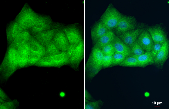
Cytokeratin 8 antibody detects Cytokeratin 8 protein at cytoplasm by immunofluorescent analysis. Sample: MDCK cells were fixed in 4% paraformaldehyde at RT for 15 min. Green: Cytokeratin 8 stained by Cytokeratin 8 antibody (GTX109489) diluted at 1:500.
![Immunohistochemical analysis (whole mount) of zebrafish embryo, using Cytokeratin 8 antibody [N1N3] (GTX109489) at 1:200 dilution.](https://www.genetex.com/upload/website/prouct_img/normal/GTX109489/GTX109489_40009_IHC-Wm_Z_22111423_167.webp "Immunohistochemical analysis (whole mount) of zebrafish embryo, using Cytokeratin 8 antibody [N1N3] (GTX109489) at 1:200 dilution.")
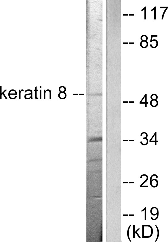
 was separated by 10% SDS-PAGE, and the membrane was blotted with Cytokeratin 8 antibody (GTX109489) diluted at 1:1000. The HRP-conjugated anti-rabbit IgG antibody (GTX213110-01) was used to detect the primary antibody.")
 A:MOLT4 (GTX27912) B:Raji (GTX27908) 10% SDS PAGE GTX109489 diluted at 1:1000")
 antibody at 1:200 dilution.")
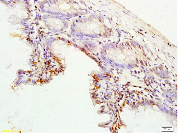
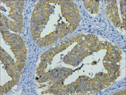
![Cytokeratin 8 antibody [N1N3] detects Cytokeratin 8 protein at cytoplasm and membrane in mouse duodenum by immunohistochemical analysis. Sample: Paraffin-embedded mouse duodenum. Cytokeratin 8 antibody [N1N3] (GTX109489) diluted at 1:500.
Antigen Retrieval: Citrate buffer, pH 6.0, 15 min](https://www.genetex.com/upload/website/prouct_img/normal/GTX109489/GTX109489_40009_20150417_IHC_M_w_23060500_722.webp "Cytokeratin 8 antibody [N1N3] detects Cytokeratin 8 protein at cytoplasm and membrane in mouse duodenum by immunohistochemical analysis. Sample: Paraffin-embedded mouse duodenum. Cytokeratin 8 antibody [N1N3] (GTX109489) diluted at 1:500.
Antigen Retrieval: Citrate buffer, pH 6.0, 15 min")
 antibody at 1:500 dilution.
Antigen Retrieval: Citrate buffer, pH 6.0, 15 min")
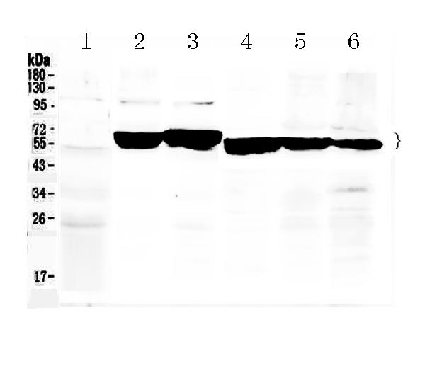
![Rat tissue extract (50 μg) was separated by 10% SDS-PAGE, and the membrane was blotted with Cytokeratin 8 antibody [N1N3] (GTX109489) diluted at 1:1000. The HRP-conjugated anti-rabbit IgG antibody (GTX213110-01) was used to detect the primary antibody.](https://www.genetex.com/upload/website/prouct_img/normal/GTX109489/GTX109489_40009_20250228_WB_R_intestine_25030600_467.webp "Rat tissue extract (50 μg) was separated by 10% SDS-PAGE, and the membrane was blotted with Cytokeratin 8 antibody [N1N3] (GTX109489) diluted at 1:1000. The HRP-conjugated anti-rabbit IgG antibody (GTX213110-01) was used to detect the primary antibody.")
Cytokeratin 8 antibody detects Cytokeratin 8 protein at cytoplasm by immunofluorescent analysis. Sample: MDCK cells were fixed in 4% paraformaldehyde at RT for 15 min. Green: Cytokeratin 8 stained by Cytokeratin 8 antibody (GTX109489) diluted at 1:500.
Cytokeratin 8 antibody [N1N3]
GTX109489
ApplicationsImmunoFluorescence, Western Blot, ImmunoCytoChemistry, ImmunoHistoChemistry, ImmunoHistoChemistry Paraffin
Product group Antibodies
ReactivityCanine, Human, Mouse, Rat, Zebra Fish
TargetKRT8
Overview
- SupplierGeneTex
- Product NameCytokeratin 8 antibody [N1N3]
- Delivery Days Customer9
- Application Supplier NoteWB: 1:500-1:3000. ICC/IF: 1:100-1:1000. IHC-P: 1:100-1:1000. *Optimal dilutions/concentrations should be determined by the researcher.Not tested in other applications.
- ApplicationsImmunoFluorescence, Western Blot, ImmunoCytoChemistry, ImmunoHistoChemistry, ImmunoHistoChemistry Paraffin
- CertificationResearch Use Only
- ClonalityPolyclonal
- Concentration0.53 mg/ml
- ConjugateUnconjugated
- Gene ID3856
- Target nameKRT8
- Target descriptionkeratin 8
- Target synonymsCARD2, CK-8, CK8, CYK8, K2C8, K8, KO, keratin, type II cytoskeletal 8, cytokeratin-8, keratin 8, type II, type-II keratin Kb8
- HostRabbit
- IsotypeIgG
- Protein IDP05787
- Protein NameKeratin, type II cytoskeletal 8
- Scientific DescriptionThis gene is a member of the type II keratin family clustered on the long arm of chromosome 12. Type I and type II keratins heteropolymerize to form intermediate-sized filaments in the cytoplasm of epithelial cells. The product of this gene typically dimerizes with keratin 18 to form an intermediate filament in simple single-layered epithelial cells. This protein plays a role in maintaining cellular structural integrity and also functions in signal transduction and cellular differentiation. Mutations in this gene cause cryptogenic cirrhosis. [provided by RefSeq]
- ReactivityCanine, Human, Mouse, Rat, Zebra Fish
- Storage Instruction-20°C or -80°C,2°C to 8°C
- UNSPSC41116161
Datasheet
Related products
Product group Antibodies
Anti-Keratin 8 AntibodyA95180
ApplicationsWestern Blot, ELISA, ImmunoHistoChemistry
ReactivityHuman, Mouse, Rat
- SizePrice
Product group Antibodies

Anti-Human Cytokeratin 8 [1E8-C6-B4]Ab02687-1.1
ApplicationsImmunoFluorescence, ImmunoPrecipitation, Western Blot
ReactivityHuman
TargetKRT8
- SizePrice
Product group Antibodies
Anti-Cytokeratin 8/KRT8 Antibody Picoband(r)A01421-CARRIER-FREE
ApplicationsWestern Blot, ImmunoHistoChemistry
ReactivityHuman, Mouse, Rat
TargetKRT8
- SizePrice
Product group Antibodies
References
ApplicationsFlow Cytometry, ImmunoFluorescence, Western Blot, ELISA, ImmunoCytoChemistry, ImmunoHistoChemistry, ImmunoHistoChemistry Frozen, ImmunoHistoChemistry Paraffin
TargetKRT8
- SizePrice
Product group Antibodies
KRT8 Monoclonal AntibodyCSB-MA000212
ApplicationsWestern Blot, ELISA, ImmunoHistoChemistry
ReactivityHuman, Mouse, Rat
TargetKRT8
- SizePrice
Product group Antibodies
Krt8 Polyclonal AntibodyCAC07095
ApplicationsImmunoFluorescence, Western Blot, ELISA, ImmunoHistoChemistry
TargetKRT8
- SizePrice
![IHC-P analysis of human skin tissue using GTX17139 Cytokeratin 8 + 18 antibody [5D3].](https://www.genetex.com/upload/website/prouct_img/normal/GTX17139/GTX17139_20191203_IHC-P_28_w_23060620_418.webp)
Product group Antibodies
References
ApplicationsImmunoHistoChemistry, ImmunoHistoChemistry Paraffin
ReactivityHuman
TargetKRT8
- SizePrice
![IHC-P analysis of human colon tissue using GTX20758 Cytokeratin 8 antibody [35bH11].](https://www.genetex.com/upload/website/prouct_img/normal/GTX20758/GTX20758_20191203_IHC-P_45_w_23060620_206.webp)
Product group Antibodies
ApplicationsImmunoHistoChemistry, ImmunoHistoChemistry Paraffin
ReactivityHuman
TargetKRT8
- SizePrice