

Cytokeratin 8/18(C-51), Biotin conjugate, 0.1mg/mL [26628-22-8]
BNCB0034
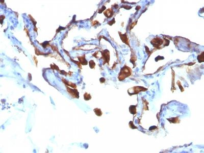
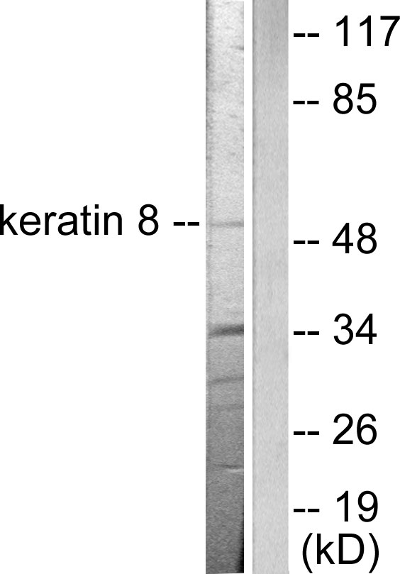
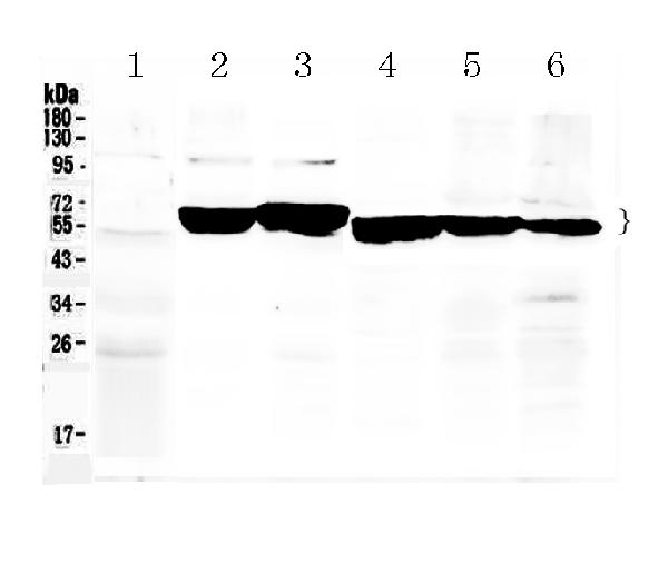
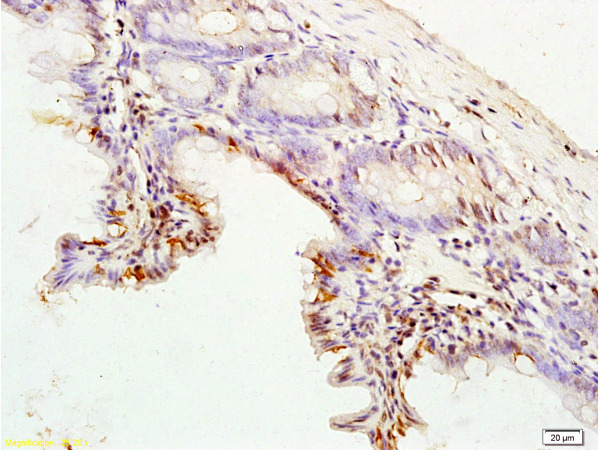
ApplicationsImmunoFluorescence, Western Blot, ImmunoHistoChemistry, ImmunoHistoChemistry Paraffin
Product group Antibodies
ReactivityBovine, Human, Monkey, Mouse, Porcine, Sheep
TargetKRT8
Overview
- SupplierBiotium
- Product NameCytokeratin 8/18(C-51), Biotin conjugate, 0.1mg/mL [26628-22-8]
- Delivery Days Customer9
- ApplicationsImmunoFluorescence, Western Blot, ImmunoHistoChemistry, ImmunoHistoChemistry Paraffin
- CAS Number26628-22-8
- CertificationResearch Use Only
- ClonalityMonoclonal
- Clone IDC-51
- Concentration0.1 mg/ml
- ConjugateBiotin
- Gene ID3856
- Target nameKRT8
- Target descriptionkeratin 8
- Target synonymsCARD2, CK-8, CK8, CYK8, K2C8, K8, KO, keratin, type II cytoskeletal 8, cytokeratin-8, keratin 8, type II, type-II keratin Kb8
- HostMouse
- IsotypeIgG1
- Protein IDP05783
- Protein NameKeratin, type I cytoskeletal 18
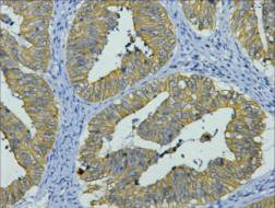
- Scientific DescriptionCytokeratin 8 (CK8) belongs to the type II (or B or basic) subfamily of high molecular weight cytokeratins and exists in combination with cytokeratin 18 (CK18). This MAb recognizes all simple epithelia including glandular epithelium, for example thyroid, female breast, gastrointestinal tract, respiratory tract, and urogenital tract including transitional epithelium. All adenocarcinomas and most squamous carcinomas are positive but keratinizing squamous carcinomas are usually negative. This antibody is useful in demonstrating the presence of Paget cells; there is very little keratin 18 in the normal epidermis so only Paget cells are stained.Primary antibodies are available purified, or with a selection of fluorescent CF® Dyes and other labels. CF® Dyes offer exceptional brightness and photostability. Note: Conjugates of blue fluorescent dyes like CF®405S and CF®405M are not recommended for detecting low abundance targets, because blue dyes have lower fluorescence and can give higher non-specific background than other dye colors.
- SourceAnimal
- ReactivityBovine, Human, Monkey, Mouse, Porcine, Sheep
- Storage Instruction2°C to 8°C,RT
- UNSPSC41116161
MSDS
Related products
Product group Antibodies
Anti-Keratin 8 AntibodyA95180
ApplicationsWestern Blot, ELISA, ImmunoHistoChemistry
ReactivityHuman, Mouse, Rat
- SizePrice
Product group Antibodies

Anti-Human Cytokeratin 8 [1E8-C6-B4]Ab02687-1.1
ApplicationsImmunoFluorescence, ImmunoPrecipitation, Western Blot
ReactivityHuman
TargetKRT8
- SizePrice
Product group Antibodies
Anti-Cytokeratin 8/KRT8 Antibody Picoband(r)A01421-CARRIER-FREE
ApplicationsWestern Blot, ImmunoHistoChemistry
ReactivityHuman, Mouse, Rat
TargetKRT8
- SizePrice
Product group Antibodies
References
ApplicationsFlow Cytometry, ImmunoFluorescence, Western Blot, ELISA, ImmunoCytoChemistry, ImmunoHistoChemistry, ImmunoHistoChemistry Frozen, ImmunoHistoChemistry Paraffin
TargetKRT8
- SizePrice
Product group Antibodies
KRT8 Monoclonal AntibodyCSB-MA000212
ApplicationsWestern Blot, ELISA, ImmunoHistoChemistry
ReactivityHuman, Mouse, Rat
TargetKRT8
- SizePrice
Product group Antibodies
Krt8 Polyclonal AntibodyCAC07095
ApplicationsImmunoFluorescence, Western Blot, ELISA, ImmunoHistoChemistry
TargetKRT8
- SizePrice
Product group Antibodies

ApplicationsWestern Blot, ELISA
ReactivityHuman
TargetKRT8
- SizePrice

Product group Antibodies
Anti-KRT8-25ulHPA049866
ApplicationsWestern Blot, ImmunoCytoChemistry, ImmunoHistoChemistry
ReactivityHuman
- SizePrice