

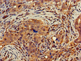
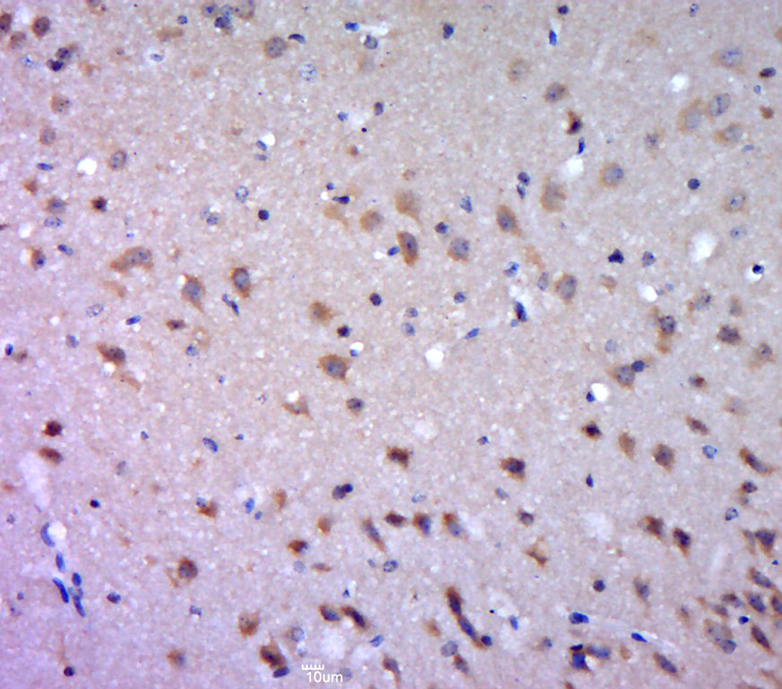
Immunohistochemistry of paraffin-embedded human pancreatic cancer using CSB-PA006817LA01HU at dilution of 1:100
Immunohistochemistry of paraffin-embedded human pancreatic cancer using CSB-PA006817LA01HU at dilution of 1:100
DGAT1 Antibody
CSB-PA006817LA01HU
ApplicationsELISA, ImmunoHistoChemistry
Product group Antibodies
ReactivityHuman
TargetDGAT1
Overview
- SupplierCusabio
- Product NameDGAT1 Antibody
- Delivery Days Customer20
- ApplicationsELISA, ImmunoHistoChemistry
- CertificationResearch Use Only
- ClonalityPolyclonal
- ConjugateUnconjugated
- Gene ID8694
- Target nameDGAT1
- Target descriptiondiacylglycerol O-acyltransferase 1
- Target synonymsARAT, ARGP1, DGAT, DIAR7, diacylglycerol O-acyltransferase 1, ACAT related gene product 1, acyl coenzyme A:cholesterol acyltransferase related gene 1, acyl-CoA retinol O-fatty-acyltransferase, acyl-CoA:diacylglycerol acyltransferase, diglyceride acyltransferase
- HostRabbit
- IsotypeIgG
- Protein IDO75907
- Protein NameDiacylglycerol O-acyltransferase 1
- Scientific DescriptionCatalyzes the terminal and only committed step in triacylglycerol synthesis by using diacylglycerol and fatty acyl CoA as substrates. In contrast to DGAT2 it is not essential for survival. May be involved in VLDL (very low density lipoprotein) assembly. In liver, plays a role in esterifying exogenous fatty acids to glycerol. Functions as the major acyl-CoA retinol acyltransferase (ARAT) in the skin, where it acts to maintain retinoid homeostasis and prevent retinoid toxicity leading to skin and hair disorders.
- ReactivityHuman
- Storage Instruction-20°C or -80°C
- UNSPSC41116161
Related products
Product group Antibodies
Anti-DGAT1 AntibodyA12028
ApplicationsImmunoFluorescence, Western Blot, ImmunoCytoChemistry
ReactivityHuman, Mouse, Rat
- SizePrice
Product group Antibodies

DGAT1 Antibody (aa1-90)LS-C750310
ApplicationsWestern Blot
ReactivityHuman, Mouse, Rat
TargetDGAT1
- SizePrice
Product group Antibodies

Goat anti-DGAT1EB07575
ApplicationsFlow Cytometry, ImmunoFluorescence, Western Blot, ELISA, ImmunoHistoChemistry
ReactivityBovine, Canine, Human, Mouse, Rat
TargetDGAT1
- SizePrice
Product group Antibodies
References
DGAT1 Polyclonal AntibodyBS-2332R
ApplicationsImmunoFluorescence, Western Blot, ELISA, ImmunoCytoChemistry, ImmunoHistoChemistry, ImmunoHistoChemistry Frozen, ImmunoHistoChemistry Paraffin
ReactivityCanine, Equine, Human, Mouse, Porcine, Rat
TargetDGAT1
- SizePrice
Product group Antibodies
ApplicationsImmunoPrecipitation, Western Blot, ImmunoCytoChemistry, ImmunoHistoChemistry
TargetDGAT1
- SizePrice

Product group Antibodies
Anti-DGAT1 Antibody Picoband(r)PB9801-CARRIER-FREE
ApplicationsWestern Blot
ReactivityHuman, Rat
TargetDGAT1
- SizePrice
Product group Antibodies
References
DGAT1 antibodyGTX48577
ApplicationsImmunoFluorescence, Western Blot, ImmunoCytoChemistry
ReactivityBovine, Goat, Human, Mouse, Primate, Rat, Sheep, Zebra Fish
TargetDGAT1
- SizePrice
Product group Antibodies
Anti-DGAT1 Antibody144-06857
ApplicationsWestern Blot, ImmunoHistoChemistry
ReactivityHuman, Mouse, Rat
TargetDGAT1
- SizePrice