

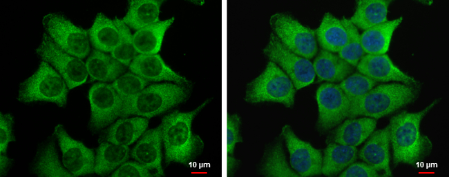
Dvl-2 antibody [N1N3] detects Dvl-2 protein at cytoplasm by immunofluorescent analysis. Sample: HCT 116 cells were fixed in 4% paraformaldehyde at RT for 15 min. Green: Dvl-2 protein stained by Dvl-2 antibody [N1N3] (GTX111156) diluted at 1:500. Blue: Hoechst 33342 staining.
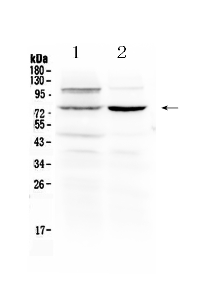
![Various whole cell extracts (30 μg) were separated by 7.5% SDS-PAGE, and the membrane was blotted with Dishevelled 2 antibody [N1N3] (GTX111156) diluted at 1:5000. The HRP-conjugated anti-rabbit IgG antibody (GTX213110-01) was used to detect the primary antibody.](https://www.genetex.com/upload/website/prouct_img/normal/GTX111156/GTX111156_40079_20170518_WB_M_w_23060500_628.webp "Various whole cell extracts (30 μg) were separated by 7.5% SDS-PAGE, and the membrane was blotted with Dishevelled 2 antibody [N1N3] (GTX111156) diluted at 1:5000. The HRP-conjugated anti-rabbit IgG antibody (GTX213110-01) was used to detect the primary antibody.")
![Various whole cell extracts (30 μg) were separated by 7.5% SDS-PAGE, and the membrane was blotted with Dishevelled 2 antibody [N1N3] (GTX111156) diluted at 1:5000. The HRP-conjugated anti-rabbit IgG antibody (GTX213110-01) was used to detect the primary antibody.](https://www.genetex.com/upload/website/prouct_img/normal/GTX111156/GTX111156_40079_20170518_WB_R_w_23060500_486.webp "Various whole cell extracts (30 μg) were separated by 7.5% SDS-PAGE, and the membrane was blotted with Dishevelled 2 antibody [N1N3] (GTX111156) diluted at 1:5000. The HRP-conjugated anti-rabbit IgG antibody (GTX213110-01) was used to detect the primary antibody.")
![Various whole cell extracts (30 μg) were separated by 7.5% SDS-PAGE, and the membranes were blotted with Dishevelled 2 antibody [N1N3] (GTX111156) diluted at 1:5000 and competitor's antibody (SC-13974) diluted by 1:200.](https://www.genetex.com/upload/website/prouct_img/normal/GTX111156/GTX111156_40079_20161124_WB_competitor_watermark_w_23060500_639.webp "Various whole cell extracts (30 μg) were separated by 7.5% SDS-PAGE, and the membranes were blotted with Dishevelled 2 antibody [N1N3] (GTX111156) diluted at 1:5000 and competitor's antibody (SC-13974) diluted by 1:200.")
![Various whole cell extracts (30 μg) were separated by 7.5% SDS-PAGE, and the membranes were blotted with Dishevelled 2 antibody [N1N3] (GTX111156) diluted at 1:10000 and competitor's antibody (sc-13974) diluted at 1:1000. The HRP-conjugated anti-rabbit IgG antibody (GTX213110-01) was used to detect the primary antibody.](https://www.genetex.com/upload/website/prouct_img/normal/GTX111156/GTX111156_40079_20170804_WB_competitor_watermark_w_23060500_855.webp "Various whole cell extracts (30 μg) were separated by 7.5% SDS-PAGE, and the membranes were blotted with Dishevelled 2 antibody [N1N3] (GTX111156) diluted at 1:10000 and competitor's antibody (sc-13974) diluted at 1:1000. The HRP-conjugated anti-rabbit IgG antibody (GTX213110-01) was used to detect the primary antibody.")
Dvl-2 antibody [N1N3] detects Dvl-2 protein at cytoplasm by immunofluorescent analysis. Sample: HCT 116 cells were fixed in 4% paraformaldehyde at RT for 15 min. Green: Dvl-2 protein stained by Dvl-2 antibody [N1N3] (GTX111156) diluted at 1:500. Blue: Hoechst 33342 staining.
Dishevelled 2 antibody [N1N3]
GTX111156
ApplicationsImmunoFluorescence, Western Blot, ImmunoCytoChemistry
Product group Antibodies
ReactivityHuman, Mouse, Rat
TargetDVL2
Overview
- SupplierGeneTex
- Product NameDishevelled 2 antibody [N1N3]
- Delivery Days Customer9
- Application Supplier NoteWB: 1:1000-1:20000. ICC/IF: 1:100-1:1000. *Optimal dilutions/concentrations should be determined by the researcher.Not tested in other applications.
- ApplicationsImmunoFluorescence, Western Blot, ImmunoCytoChemistry
- CertificationResearch Use Only
- ClonalityPolyclonal
- Concentration1 mg/ml
- ConjugateUnconjugated
- Gene ID1856
- Target nameDVL2
- Target descriptiondishevelled segment polarity protein 2
- Target synonymssegment polarity protein dishevelled homolog DVL-2, dishevelled 2 (homologous to Drosophila dsh), dishevelled, dsh homolog 2
- HostRabbit
- IsotypeIgG
- Protein IDO14641
- Protein NameSegment polarity protein dishevelled homolog DVL-2
- Scientific DescriptionThis gene encodes a member of the dishevelled (dsh) protein family. The vertebrate dsh proteins have approximately 40% amino acid sequence similarity with Drosophila dsh. This gene encodes a 90-kD protein that undergoes posttranslational phosphorylation to form a 95-kD cytoplasmic protein, which may play a role in the signal transduction pathway mediated by multiple Wnt proteins. The mechanisms of dishevelled function in Wnt signaling are likely to be conserved among metazoans. [provided by RefSeq]
- ReactivityHuman, Mouse, Rat
- Storage Instruction-20°C or -80°C,2°C to 8°C
- UNSPSC41116161
Datasheet
Related products
Product group Antibodies
ApplicationsImmunoFluorescence, ImmunoCytoChemistry
ReactivityMouse
- SizePrice
Product group Antibodies
Anti-Dishevelled 2/DVL2 Antibody Picoband(r)A02404-3-CARRIER-FREE
ApplicationsWestern Blot
ReactivityHuman, Mouse, Rat
TargetDVL2
- SizePrice
Product group Antibodies
Anti-DVL2 Antibody144-66425
ApplicationsImmunoFluorescence, Western Blot, ImmunoHistoChemistry
ReactivityHuman, Mouse, Rat
TargetDVL2
- SizePrice
Product group Antibodies

Anti-DVL2 [RAB-T7]Ab01770-1.1
ApplicationsImmunoFluorescence, ImmunoPrecipitation
ReactivityHuman
TargetDVL2
- SizePrice
Product group Antibodies

DVL2 / Dishevelled 2 AntibodyLS-C749710
ApplicationsImmunoFluorescence, ImmunoHistoChemistry
ReactivityHuman, Mouse, Rat
TargetDVL2
- SizePrice
Product group Antibodies
Dishevelled 2 Polyclonal AntibodyBS-10400R
ApplicationsImmunoFluorescence, Western Blot, ELISA, ImmunoCytoChemistry, ImmunoHistoChemistry, ImmunoHistoChemistry Frozen, ImmunoHistoChemistry Paraffin
ReactivityBovine, Canine, Equine, Human, Mouse, Porcine, Rat, Sheep
TargetDVL2
- SizePrice
Product group Antibodies
DVL2 AntibodyCSB-PA007286ESR1HU
ApplicationsImmunoFluorescence, Western Blot, ELISA, ImmunoHistoChemistry
ReactivityHuman
TargetDVL2
- SizePrice
Product group Antibodies
Dvl2 Polyclonal AntibodyCAC10639
ApplicationsImmunoFluorescence, Western Blot, ELISA, ImmunoHistoChemistry
TargetDVL2
- SizePrice
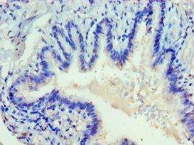
![cytoplasm in the photoreceptor Immunohistochemistry of flat-mounted zebrafish retina using an antibody against Dishevelled 2 antibody [N2C2], Internal (GTX103878) at a 1:200 dilution.](https://www.genetex.com/upload/website/prouct_img/normal/GTX103878/GTX103878_39918_IHC_Z_22111423_689.webp)
Product group Antibodies
ApplicationsWestern Blot, ImmunoHistoChemistry, ImmunoHistoChemistry Frozen, ImmunoHistoChemistry Paraffin
ReactivityHuman, Mouse, Zebra Fish
TargetDVL2
- SizePrice

Product group Antibodies
Anti-DVL2 AntibodyHPA021611
ApplicationsWestern Blot, ImmunoCytoChemistry
ReactivityHuman
TargetDVL2
- SizePrice