

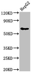
DLAT Polyclonal Antibody
RD83712A
ApplicationsImmunoFluorescence
Product group Antibodies
ReactivityHuman, Mouse, Rat
TargetDLAT
Overview
- SupplierReddot Biotech
- Product NameDLAT Polyclonal Antibody
- Delivery Days Customer5
- ApplicationsImmunoFluorescence
- CertificationResearch Use Only
- Concentration1 mg/ml
- ConjugateUnconjugated
- Gene ID1737
- Target nameDLAT
- Target descriptiondihydrolipoamide S-acetyltransferase
- Target synonymsDLTA, E2, PBC, PDC-E2, PDCE2, dihydrolipoyllysine-residue acetyltransferase component of pyruvate dehydrogenase complex, mitochondrial, 70 kDa mitochondrial autoantigen of primary biliary cirrhosis, E2 component of pyruvate dehydrogenase complex, M2 antigen complex 70 kDa subunit, dihydrolipoamide acetyltransferase component of pyruvate dehydrogenase complex, pyruvate dehydrogenase complex component E2
- HostRabbit
- IsotypeIgG
- Scientific DescriptionThis gene encodes component E2 of the multi-enzyme pyruvate dehydrogenase complex (PDC). PDC resides in the inner mitochondrial membrane and catalyzes the conversion of pyruvate to acetyl coenzyme A. The protein product of this gene, dihydrolipoamide acetyltransferase, accepts acetyl groups formed by the oxidative decarboxylation of pyruvate and transfers them to coenzyme A. Dihydrolipoamide acetyltransferase is the antigen for antimitochondrial antibodies. These autoantibodies are present in nearly 95% of patients with the autoimmune liver disease primary biliary cirrhosis (PBC). In PBC, activated T lymphocytes attack and destroy epithelial cells in the bile duct where this protein is abnormally distributed and overexpressed. PBC enventually leads to cirrhosis and liver failure. Mutations in this gene are also a cause of pyruvate dehydrogenase E2 deficiency which causes primary lactic acidosis in infancy and early childhood.
- ReactivityHuman, Mouse, Rat
- Storage Instruction-20°C
- UNSPSC12352203
Related products
Product group Antibodies
Anti-DLAT Antibody144-60697
ApplicationsWestern Blot, ImmunoHistoChemistry
ReactivityHuman, Mouse, Rat
TargetDLAT
- SizePrice
Product group Antibodies
DLAT Polyclonal AntibodyCAC12898
ApplicationsImmunoPrecipitation, Western Blot, ELISA
TargetDLAT
- SizePrice
Product group Antibodies
DLAT AntibodyCSB-PA006926LA01HU
ApplicationsImmunoPrecipitation, Western Blot, ELISA
ReactivityHuman
TargetDLAT
- SizePrice
Product group Antibodies

DLAT / PDC-E2 AntibodyLS-C766087
ApplicationsWestern Blot, ELISA, ImmunoHistoChemistry
ReactivityHuman, Mouse, Rat
TargetDLAT
- SizePrice

Product group Antibodies
Anti-DLAT AntibodyA29546
ApplicationsWestern Blot, ImmunoHistoChemistry
ReactivityHuman, Mouse, Rat
- SizePrice
Product group Antibodies
DLAT Recombinant Antibody, AbBy Fluor-405 ConjugatedBSM-62155R-BF405
ApplicationsFlow Cytometry, ImmunoFluorescence, Western Blot, ImmunoCytoChemistry
ReactivityHuman, Mouse, Rat
TargetDLAT
- SizePrice

Product group Antibodies
Anti-DLAT AntibodyHPA040786
ApplicationsWestern Blot, ImmunoCytoChemistry, ImmunoHistoChemistry
ReactivityHuman
TargetDLAT
- SizePrice
Product group Antibodies
TargetDLAT
- SizePrice
Product group Antibodies
References
DLAT antibodyGTX109766
ApplicationsImmunoFluorescence, Western Blot, ImmunoCytoChemistry, ImmunoHistoChemistry, ImmunoHistoChemistry Paraffin
ReactivityHuman
TargetDLAT
- SizePrice