

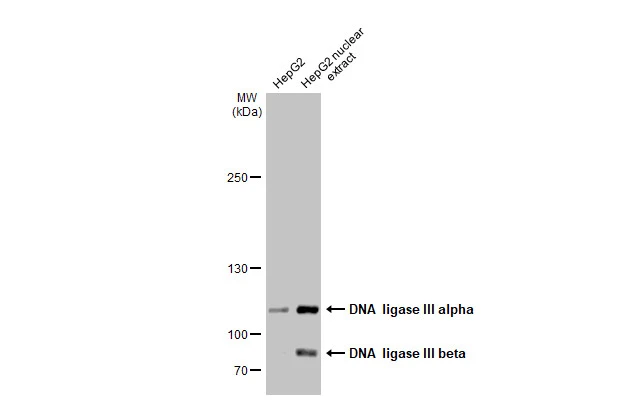
HepG2 whole cell and nuclear extracts (30 μg) were separated by 5% SDS-PAGE, and the membrane was blotted with DNA ligase III antibody [C2C3], C-term (GTX103197) diluted at 1:500. The HRP-conjugated anti-rabbit IgG antibody (GTX213110-01) was used to detect the primary antibody.
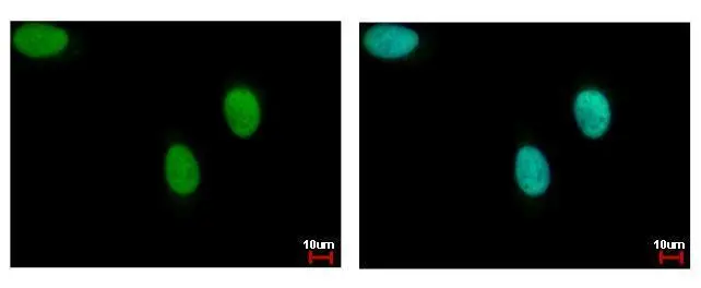
![DNA ligase III antibody [C2C3], C-term detects DNA ligase III protein at nucleus by immunofluorescent analysis. Sample: HeLa cells were fixed in 4% paraformaldehyde at RT for 15 min. Green: DNA ligase III protein stained by DNA ligase III antibody [C2C3], C-term (GTX103197) diluted at 1:200. Red: phalloidin, a cytoskeleton marker, diluted at 1:200. Scale bar = 10 μm.](https://www.genetex.com/upload/website/prouct_img/normal/GTX103197/GTX103197_40142_20160113_IFA_w_23060119_768.webp "DNA ligase III antibody [C2C3], C-term detects DNA ligase III protein at nucleus by immunofluorescent analysis. Sample: HeLa cells were fixed in 4% paraformaldehyde at RT for 15 min. Green: DNA ligase III protein stained by DNA ligase III antibody [C2C3], C-term (GTX103197) diluted at 1:200. Red: phalloidin, a cytoskeleton marker, diluted at 1:200. Scale bar = 10 μm.")
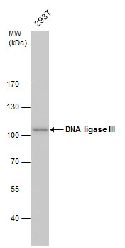
![Non-transfected (–) and transfected (+) 293T whole cell extracts (30 μg) were separated by 7.5% SDS-PAGE, and the membrane was blotted with DNA ligase III antibody [C2C3], C-term (GTX103197) diluted at 1:500.](https://www.genetex.com/upload/website/prouct_img/normal/GTX103197/GTX103197_40142_20160908_WB_shRNA_watermark_w_23060119_644.webp "Non-transfected (–) and transfected (+) 293T whole cell extracts (30 μg) were separated by 7.5% SDS-PAGE, and the membrane was blotted with DNA ligase III antibody [C2C3], C-term (GTX103197) diluted at 1:500.")
HepG2 whole cell and nuclear extracts (30 μg) were separated by 5% SDS-PAGE, and the membrane was blotted with DNA ligase III antibody [C2C3], C-term (GTX103197) diluted at 1:500. The HRP-conjugated anti-rabbit IgG antibody (GTX213110-01) was used to detect the primary antibody.
DNA ligase III antibody [C2C3], C-term
GTX103197
ApplicationsImmunoFluorescence, Western Blot, ImmunoCytoChemistry
Product group Antibodies
ReactivityHamster, Human
TargetLIG3
Overview
- SupplierGeneTex
- Product NameDNA ligase III antibody [C2C3], C-term
- Delivery Days Customer9
- Application Supplier NoteWB: 1:500-1:3000. ICC/IF: 1:100-1:1000. *Optimal dilutions/concentrations should be determined by the researcher.Not tested in other applications.
- ApplicationsImmunoFluorescence, Western Blot, ImmunoCytoChemistry
- CertificationResearch Use Only
- ClonalityPolyclonal
- Concentration0.15 mg/ml
- ConjugateUnconjugated
- Gene ID3980
- Target nameLIG3
- Target descriptionDNA ligase 3
- Target synonymsLIG2, LIG3alpha, MTDPS20, DNA ligase 3, ligase II, DNA, ATP-dependent, ligase III, DNA, ATP-dependent, polydeoxyribonucleotide synthase [ATP] 3
- HostRabbit
- IsotypeIgG
- Protein IDP49916
- Protein NameDNA ligase 3
- Scientific DescriptionThis gene is a member of the DNA ligase family. Each member of this family encodes a protein that catalyzes the joining of DNA ends but they each have a distinct role in DNA metabolism. The protein encoded by this gene is involved in excision repair and is located in both the mitochondria and nucleus, with translation initiation from the upstream start codon allowing for transport to the mitochondria and translation initiation from a downstream start codon allowing for transport to the nucleus. Additionally, alternate transcriptional splice variants, encoding different isoforms, have been characterized. [provided by RefSeq]
- ReactivityHamster, Human
- Storage Instruction-20°C or -80°C,2°C to 8°C
- UNSPSC41116161
Datasheet
Related products
Product group Antibodies
LIG3 AntibodyCSB-PA002166
ApplicationsWestern Blot, ELISA
ReactivityHuman, Mouse, Rat
TargetLIG3
- SizePrice
Product group Antibodies
Anti-LIG3 AntibodyA29907
ApplicationsImmunoFluorescence, Western Blot, ImmunoHistoChemistry
ReactivityHuman, Mouse, Rat
- SizePrice

Product group Antibodies
Anti-DNA Ligase III/LIG3 Antibody Picoband(r)A02741-2-CARRIER-FREE
ApplicationsFlow Cytometry, Western Blot, ELISA
ReactivityHuman, Mouse, Rat
TargetLIG3
- SizePrice
Product group Antibodies

LIG3 / DNA Ligase III AntibodyLS-C748557
ApplicationsWestern Blot, ImmunoHistoChemistry
ReactivityHuman, Mouse, Rat
TargetLIG3
- SizePrice
![HepG2 whole cell and nuclear extracts (30 μg) were separated by 7.5% SDS-PAGE, and the membrane was blotted with DNA ligase III antibody [HL2280] (GTX638333) diluted at 1:1000. The HRP-conjugated anti-rabbit IgG antibody (GTX213110-01) was used to detect the primary antibody.](https://www.genetex.com/upload/website/prouct_img/normal/GTX638333/GTX638333_T-44977_20230317_WB_Fraction_23032022_926.webp)
Product group Antibodies
DNA ligase III antibody [HL2280]GTX638333
ApplicationsImmunoFluorescence, Western Blot, ImmunoCytoChemistry, ImmunoHistoChemistry, ImmunoHistoChemistry Paraffin
ReactivityHuman, Mouse, Rat
TargetLIG3
- SizePrice
Product group Antibodies
DNA ligase III antibody [1F3]GTX70143
ApplicationsImmunoFluorescence, ImmunoPrecipitation, Western Blot, ImmunoCytoChemistry, Neutralisation/Blocking, Other Application
ReactivityChicken, Human, Mouse
TargetLIG3
- SizePrice
Product group Antibodies
DNA ligase III antibodyGTX103172
ApplicationsImmunoFluorescence, Western Blot, ImmunoCytoChemistry, ImmunoHistoChemistry, ImmunoHistoChemistry Paraffin
ReactivityHuman, Mouse
TargetLIG3
- SizePrice
Product group Antibodies
Anti-LIG3 Antibody144-60365
ApplicationsWestern Blot, ImmunoHistoChemistry
ReactivityHuman, Mouse, Rat
TargetLIG3
- SizePrice